This test is performed at the beginning of pregnancy to identify possible genetic pathologies in the fetus. The first screening includes a blood test and an ultrasound examination. Only in combination they give an accurate result. How to prepare for the procedure, to whom it is indicated, and is it possible to refuse it?
What is pregnancy screening
This is an extremely important examination carried out when carrying a child. It allows you to assess the condition and development of an unborn baby. When prescribing screening, the doctor takes into account the characteristics of the mother's body (weight, height, bad habits, chronic diseases), which may affect the test results.
With ultrasound, the doctor examines the development of the physique of the fetus and determines if there are any pathologies. If violations are found, treatment can be started on time.
When is the first screening done?
Patients are interested in when they do the 1st screening, and if there is a time frame to delay or accelerate testing. The timing is set by the gynecologist leading the pregnancy. It is often assigned 10 to 13 weeks after conception. Despite the short duration of pregnancy, tests accurately show the presence of chromosomal disorders in the fetus.
Be sure to screen women at risk by week 13:
- over 35 years of age;
- under the age of 18;
- having genetic diseases in the family;
- survivors of a spontaneous abortion;
- who gave birth to children with genetic disorders;
- sick with an infectious disease after conception;
- who conceived a child from a relative.
Screening is prescribed for women who have had viral diseases in the first trimester. Often, not knowing what is in position, a pregnant woman is treated with conventional drugs that negatively affect the development of the embryo.
What should show
Thanks to the first screening, the expectant mother and the doctor will know exactly how the baby is developing and whether he is healthy.
Biochemical analysis of the 1st screening during pregnancy has certain indicators:
- HCG norm- detects Edwards syndrome when the indicators are below the established ones. If they are overestimated, then the development of Down syndrome is suspected.
- Plasma protein (PAPP-A), the value of which is below the established norms indicates the tendency of the fetus to diseases in the future.
An ultrasound examination should show:
- how the fetus is located to eliminate the risk of ectopic pregnancy;
- what kind of pregnancy: multiple or singleton;
- whether the fetal heartbeat corresponds to the norms of development;
- embryo length, head circumference, limb length;
- the presence of external defects and violations of internal organs;
- collar space thickness. With healthy development, it corresponds to 2 cm. If there is a seal, then the presence of pathology is likely;
- condition of the placenta to eliminate the risk of dysfunction.
| Diagnostics | Gestation terms | Indicators | Meaning |
Ultrasound examination of the fetus. Depending on the intrauterine location and carry out: - through the skin - transvaginally. | 10 to 14 weeks | The coccyx-parietal size shows the maximum distance from the back of the head to the coccyx of the fetus. | Allows you to accurately determine the duration of pregnancy, and confirm the presence of pathology. |
| The thickness of the collar space (the neck fold in which fluid accumulates). | What matters is not the actual presence of fluid (all embryos have it), but its quantity. | ||
| Determining the length of the nasal bone. | If the nasal bone is not visualized, and the thickness of the cervical fold is increased, then the likelihood of developing Down syndrome is high. | ||
| Rhythms of heartbeats. | 147-171 beats per minute. | ||
| Biparietal head size - the distance between the extreme points of the crown in the fetal skull. | Helps to determine the presence of fetal pathologies, and confirm the calculations of the moment of conception. | ||
| Biochemical (hormonal) analysis, in which the venous blood of the expectant mother is taken in the amount of 10 ml | 10 to 13 weeks | Chorionic gonadotropin, which reveals the pathology of the placenta, Edwards syndrome and Down syndrome. | A decrease in the level of hCG in the blood during gestation or a slowdown in its growth indicates the risk of spontaneous miscarriage or the development of an ectopic pregnancy. |
| Protein A, a protein produced by the placenta. | Screening transcript of the 1st trimester is indicated in units of Mom. With MoM from 0.5 to 2.5, the indicators are considered normal. |
A comprehensive examination, the results of which are shown by the first screening, allows you to detect various genetic pathologies. If a serious illness is confirmed that threatens the quality of life and health of the unborn child, then parents are offered to terminate the pregnancy artificially.
To accurately confirm the diagnosis, a woman undergoes a biopsy and a puncture of the amniotic membrane to obtain amniotic fluid and examine it in the laboratory. Only after this can we confidently say that the pathology exists, and it is possible to make a final decision about the further course of pregnancy and the fate of the child.
Preparation and conduct of screening
The gynecologist leading the pregnancy tells the woman in detail what preparation for the procedure should be carried out. He also informs about the standard examination norms. All points of interest to her should be discussed without hiding information. There are several mandatory nuances for screening the first weeks.
- Tests for hormones are given on the same day. It is better to do the 1st screening in one laboratory. The expectant mother should not worry and understand that it is extremely necessary for her to donate blood from a vein. Unpleasant sensations during the delivery of the analysis will quickly pass, the main thing is to get the result.
- They donate blood on an empty stomach. You can drink some boiled water if you are very thirsty.
- Weighing. Before screening, it is advisable to weigh yourself, as weight and height data are important for the procedure.
The results of the tests are received by the doctor or the pregnant woman herself.
Results and norms of the study
Usually, laboratories issue forms that indicate the standard norms and the results of the pregnant woman obtained in the laboratory. The future mother will be able to easily understand them.
HCG norms at the first screening
These indicators are normal and do not indicate the presence of violations.
Indicators of ultrasound diagnostics
Based on the results, it is possible to determine the symmetry of the cerebral hemispheres of the fetus and track how the internal organs develop. But the main task of the procedure is to identify chromosomal pathologies, and eliminate the risk of their development at a later date.
So screening allows you to timely detect:
- chromosomal abnormalities (triploidy, characterized by an additional set of chromosomes);
- defects in the development of the nervous system;
- umbilical hernia;
- possible presence of Down syndrome;
- predisposition to Patau's syndrome, manifested by the receipt of an embryo of 3 thirteenth chromosomes instead of two. Most of the children born with this rare disease have a lot of physical abnormalities and die during the first years;
- de Lange syndrome, characterized by gene mutations. Such children are severely mentally retarded and have significant physical defects;
- Edwards syndrome is characterized by the presence of an extra 18th chromosome. Such children are severely retarded physically and mentally, and are more likely to be born prematurely;
- Lemli-Opitz syndrome, characterized by severe mental and physical retardation.
If an umbilical hernia is detected, a violation of the internal organs, a high heart rate, Patau syndrome is suspected. In the absence of the nasal bone or its too small size, one existing umbilical artery and a low heart rate, the threat of Edwards syndrome is noted.
When the gestational age is accurately established, but the ultrasound does not determine the nasal bone, and the facial contours are not expressed, this indicates Down's syndrome. Only an experienced specialist is engaged in deciphering the 1st screening, since erroneous results can lead to strong feelings for future parents
When to Start Worrying as a Mom-to-be
As you know, there is a human factor everywhere, and even serious laboratories can make mistakes. Incorrect results shown by biochemistry are confused with genetic defects. That happens:
- in mothers with diabetes;
- in those carrying twins;
- with early or late 1st screening;
- with an ectopic pregnancy.
False results are associated with factors such as:
- obesity of the expectant mother;
- conception through IVF, while the levels of protein A will be low;
- experiences and stressful situations that arose on the eve of the test;
- treatment with drugs, the active component of which is progesterone.
If PAPP-A at a high rate makes you alert only when the results of ultrasound are unfavorable, then a low protein content indicates such disorders as:
- freezing of the fetus;
- pathology of the primary form of the fetal nervous system;
- high probability of spontaneous abortion;
- risk of premature onset of labor;
- Rhesus conflict between mother and baby.
The blood test is 68% correct, and only in conjunction with ultrasound can one be sure of the diagnosis. If the norms of the first screening do not meet the requirements, it will be possible to dispel fears at the next test. It must be carried out in the second trimester of gestation. When the results of the 1st screening are in doubt, you can be examined in another independent laboratory. It is important to repeat the 1st screening before the 13th week of gestation.
Parents will need to consult a geneticist who will recommend further research. When a repeated study shows that the child has a predisposition to Down syndrome, this is evidenced by the thickness of the collar space and analysis for hCG and PAPP-A. If PAPP-A is higher than expected, and all other indicators correspond to the standard, then you should not worry. In medicine, there are cases when, despite the poor prognosis of the 1st and even the 2nd screening, healthy babies were born.
We continue the topic:
>> about 2nd trimester screeningRelated videos
In obstetrics, pregnancy management involves multiple examinations of the pregnant woman and the fetus. How many times is screening done? Within 9 months, a woman needs to undergo a general examination three times at different times. It is carried out to assess the basic physiological and anatomical characteristics of the fetus and determine probable pathologies.
The first trimester of pregnancy lasts 14 weeks, by the end of this period, the first scheduled examination is scheduled, consisting of a mandatory first ultrasound of the fetus and various laboratory blood tests.
Order No. 457 Min. Health R.F. from 2000 says that screening should be carried out for all pregnant women. Every woman has the opportunity to file a waiver. However, such an act can only speak of the illiteracy of the future mother and indicate a negligent attitude towards her baby.
How many risk factors are there that are considered a reason for mandatory prenatal screening? The main reason for the examination is:
- age criterion: 35+;
- the end of previous pregnancies with miscarriage or fetal fading;
- professional hazards;
- diagnosing chromosomal pathologies in the fetus in a previous pregnancy or the birth of a child with intrauterine malformations;
- diseases of an infectious nature, transferred at the beginning of pregnancy;
- taking drugs prohibited for pregnant women;
- alcoholism, drug addiction;
- diseases transmitted by heredity, both in the family of the mother and in the family of the father of the child;
- close family ties between the parents of the baby.

 Prenatal screening is very important for those women in whose family there were genetic diseases or who gave birth to a child with chromosomal abnormalities. Screening will make it possible to diagnose the pathologies of the current pregnancy, if necessary, prescribe its termination for medical reasons
 Carrying out a prenatal examination
When is the examination carried out? Primary prenatal screening is given at the end of the first trimester. The choice of time is due to the fact that by this moment most of the systems and organs of the unborn child have already been formed, and the diagnostician has the opportunity to assess the development of the fetus, to identify pathologies in a timely manner. At week 13, ultrasound diagnostics makes it possible to determine neural tube defects and gene pathologies in the embryo.
Ultrasound diagnostics as 1 stage of screening
What tests are included in the first trimester screening? Screening consists of several types of mandatory diagnostic procedures and tests. Ultrasound diagnosis of the fetus is the main diagnostic procedure of the first trimester. It is carried out in two ways: transvaginal, that is, the sensor is inserted into the vagina, or abdominal, that is, through the anterior abdominal wall.
Ultrasound screening of the 1st trimester allows you to determine the anatomical features of the child, the presence of all organs, assess their location and development. The main dimensional fetometric indicators, the characteristics of the correct development of the fetus are also evaluated, the neck fold, head girth, biparietal diameter, etc. are measured. It is mandatory to determine the size of the coccygeal-parietal size and, accordingly, the approximate height of the child at this stage of development. With the help of prenatal ultrasound diagnostics, the quality of the placental-umbilical cord blood flow is assessed, the work of the heart muscle is characterized. Including determine the thickness of the collar space in the fetus, the deviation of this indicator from normal values \u200b\u200bmay indicate a high probability of developing chromosomal pathologies.

 The first ultrasound study is indispensable in the management of pregnancy, because it gives the doctor the opportunity to take the first fetometric indicators, determine the level of fetal development, its compliance with the gestational age
 Biochemical blood test as stage 2 of screening
Biochemical analysis of blood is carried out in the laboratory. With the help of biochemical blood testing, carried out at week 13, the level of the PAPP-A protein and the hCG hormone is established. This study is also called the "double test".
The main hormone that begins to form after fertilization of the egg is human chorionic gonadotropin. When the level of the hCG hormone is low, this indicates that there is a placental pathology. The content of the hormone in the blood above the norm indicates fetal chromosomal pathologies, or indicates a multiple pregnancy.
The level of plasma protein in a woman's blood can also indicate various abnormalities in the development of the embryo. When there is a significant decrease in the PAPP-A index, this indicates possible chromosomal abnormalities and other congenital genetic defects.
If the results of the first prenatal screening reveal a high probability of the existence of the described pathological conditions, additional diagnostic procedures are mandatory. The expectant mother is sent to the procedure for studying the properties of amniotic fluid - amniocentesis. The technique makes it possible to determine the presence of chromosomal pathologies and some genes. They also do chorionobiopsy, i.e. a biopsy of the chorionic villi is performed. To carry out the test, cells that form the placenta are taken, and with their help, prenatal and hereditary diseases are determined.
Preparatory stage of screening
The first screening study needs a preliminary preparatory stage. Prenatal examination at other stages of pregnancy does not require such careful preparation and is much easier.
What foods should not be consumed before the first screening? The day before research activities, it is recommended to limit the use of foods that are potential allergens. These are chocolate, citrus fruits, seafood, individual intolerance products. Doctors do not advise eating fried and fatty foods.
The first prenatal ultrasound is usually done transvaginally. This diagnostic technique does not require special training. If, according to indications, an abdominal examination is prescribed - an ultrasound scan performed through the skin of the abdomen, then it is necessary to drink a certain amount of clean water without gas in advance to completely fill the bladder (about 500 ml.).
Preparation for a biochemical blood test is quite simple. Screening is done in the morning on an empty stomach, so you should come to the laboratory without breakfast. In addition, it is recommended to perform standard hygiene measures before examinations, without using flavored cosmetic and hygiene products.
Normal test scores
At the first prenatal screening, a number of characteristics are determined and special attention is paid to their compliance with generally accepted standard norms:
- Using an ultrasound examination, the thickness of the collar space (TVP) is assessed.
- The size of the nasal bone. This indicator, as well as the value of TVP, allows you to make a timely diagnosis of Down syndrome. Before 11 weeks, this anatomical characteristic cannot yet be assessed, and at 13 weeks, the length of the nasal bone should be at least 3 mm.
- A characteristic that depends on the degree of fetal development is the heart rate (HR). The dependence of heart rate on the week of pregnancy is shown in the table below.
- Also on ultrasound, the value of the coccyx-parietal size (KTR) is determined and the value of the biparietal size (BPR) of the child's head is calculated.

 At the first ultrasound, the doctor necessarily checks for the presence of the nasal bone, calculates the thickness of the collar zone, and also makes other fetometric measurements. This whole complex of studies and standards makes it possible to identify genetic abnormalities and developmental delays in the early stages.
 The normative values of the indicators described above are presented in the summary table:
| week of pregnancy | TVP, mm | KTR, mm | Heart rate, beats per minute | BPR, mm |
|---|---|---|---|---|
| 10 | 1,5 - 2,2 | 31 – 41 | 161 – 179 | 14 |
| 11 | 1,6 - 2,4 | 42 – 49 | 153 – 177 | 17 |
| 12 | 1,6 - 2,5 | 52 – 62 | 150 – 174 | 20 |
| 13 | 1,7 - 2,7 | 63 – 74 | 147 – 171 | 26 |
Screening of the first trimester of pregnancy includes a mandatory biochemical determination of the amount of the hCG hormone. The normal values of this indicator in the female body correspond to the following values:
In addition to the above indicators, at the first prenatal screening, based on ultrasound data, it is imperative to assess the degree of development of the systems and organs of the unborn child. Using laboratory techniques for blood tests, determine the content of glucose and protein A.
Possible pathological conditions detected by examinations
With the help of ultrasound diagnostics carried out in the first trimester of pregnancy, important information can be obtained about the possible development of chromosomal abnormalities.
Ultrasound determines the likelihood of the existence of Down syndrome, de Lange, Patau syndrome, Edwards syndrome, anomalies in the formation of the nervous system, the appearance of an umbilical hernia and such a chromosomal anomaly as triplodia.
Screening data interpretation
When deciphering the data obtained from an ultrasound examination and a blood test, the doctor compares the results with standard normal values and calculates the probability of possible deviations. To do this, the specialist determines the coefficients that show the discrepancy between the received data and some standard values. Usually, the resulting coefficient is abbreviated MoM:
- The normal value of MoM for the first 12 - 14 weeks ranges from 0.5 - 2.5. The best MoM is 1.
- The MoM value calculated for the hCG indicator below 0.5 indicates a high risk of Edwards syndrome. MoM above 2.5 is considered a sign of the development of Down syndrome.
Based on the results of the examinations, the overall probability coefficient for the existence of serious genetic pathologies is calculated. If the value of the generalized coefficient at week 13 ranges from 1:251 to 1:399, such a survey result is considered poor, and in terms of hormone levels, coefficient values below 0.5 and more than 2.5 are considered negative indicators.
The development of two fetuses, overweight women, diabetes mellitus, or other diseases of the endocrine system can affect the results of screening, here many characteristics may deviate from the normative values. Sometimes even the psychological state affects the reliability of the data obtained during the research.
Unwanted screening results should not be cause for serious distress. No matter how high the risk of developing pathology, there is the same high probability of having a healthy child.
Such an examination consists of two parts - blood donation from a vein and ultrasound. Based on them, taking into account many of your individual factors, the geneticist makes his verdict.
Screening (from the English "screening") is a concept that includes a number of activities to detect and prevent diseases. For example, screening during pregnancy provides the doctor with complete information about the various risks of pathologies and complications in the development of the child. This makes it possible to take full measures in advance to prevent diseases, including the most severe ones.
Who needs 1st trimester screening
It is very important that the following women undergo the study:
- consanguineous with the child's father
- who have had 2 or more spontaneous abortions (premature births)
- had a missed pregnancy or stillbirth
- a woman had a viral or bacterial disease during pregnancy
- have relatives suffering from genetic pathologies
- this couple already has a child with Patau, Down syndrome or other
- there was an episode of treatment with drugs that should not be used during pregnancy, even if they were prescribed for vital signs
- pregnant over 35 years
- both future parents want to check the probability of having an affected fetus.
What to look for at the first ultrasound screening during pregnancy
The first screening shows the symmetry of the cerebral hemispheres, the presence of some of its structures, which are mandatory at this time. Look at 1 screening also:
- long tubular bones, the length of the shoulder, femur, bones of the forearm and lower leg is measured
- whether the stomach and heart are in certain places
- the size of the heart and the vessels emanating from them
- belly sizes.
What pathology does this examination reveal?
The first pregnancy screening is informative in terms of detecting:
- pathology of the rudiment of the central nervous system - the neural tube
- patau syndrome
- omphalocele - umbilical hernia, when a different number of internal organs are outside the abdominal cavity, and in the hernial sac above the skin
- down syndrome
- triploidy (triple set of chromosomes instead of double)
- Edwards syndrome
- Smith-Opitz syndrome
- de Lange syndrome.
Terms of the study
Your doctor should once again scrupulously and thoroughly, depending on the date of the last menstruation, calculate at what time you should do the first study of this kind.
How to prepare for research
Screening of the first trimester is carried out in two stages:
- The first step is an ultrasound screening. If this is to be done transvaginally, no preparation is required. If in the abdominal way, then it is necessary that the bladder is full. To do this, you need to drink half a liter of water half an hour before the study. By the way, the second screening during pregnancy is carried out transabdominally, but it does not require preparation.
- Biochemical screening. This word refers to the drawing of blood from a vein.
Given the two-stage nature of the study, preparation for the first study includes:
- bladder filling – before 1 ultrasound screening
- fasting at least 4 hours before blood sampling from a vein.
- the whole previous day to deny yourself allergenic foods: citrus fruits, chocolate, seafood
- exclude completely fatty and fried foods (1-3 days before the study)
- before the study (usually blood is taken for screening for 12 weeks before 11:00) go to the toilet in the morning, then either do not urinate for 2-3 hours, or drink half a liter of water without gas an hour before the procedure. This is necessary if the study will be performed through the abdomen
- if ultrasound diagnostics is done with a vaginal probe, then preparation for 1st trimester screening will not include filling the bladder.
How the study is done
It, like the 12-week examination, consists of two stages:
- Ultrasound screening during pregnancy. It can be performed both vaginally and through the abdomen. It feels no different from an ultrasound at 12 weeks. The difference is that it is performed by sonologists, who specialize specifically in prenatal diagnostics, using high-class equipment.
- Blood sampling from a vein in the amount of 10 ml, which should be done on an empty stomach and in a specialized laboratory.
How is the screening diagnosis of the 1st trimester? First, you go through the first ultrasound during pregnancy. It is usually performed transvaginally.
To perform the study, you will need to undress below the waist, lie on the couch, bending your legs. The doctor will insert a thin special sensor in a condom very carefully into your vagina, and during the examination they will move it a little. It doesn't hurt, but you may find a small amount of spotting on the pad the next day or the next day.
How is the first transabdominal probe screening done? In this case, you either undress to the waist, or simply lift your clothes so that your stomach is exposed for examination. With such an ultrasound screening of the 1st trimester, the sensor will move along the abdomen without causing pain or discomfort.
How is the next stage of the examination carried out? With the results of the ultrasound, you go to donate blood. In the same place, you will clarify some data that are important for the correct interpretation of the results.
You will not receive results immediately, but after a few weeks. This is how the first pregnancy screening takes place.
Deciphering the results
1. Normal ultrasound data
Deciphering the first screening begins with the interpretation of ultrasound diagnostic data. Ultrasound rules:
Coccyx-parietal size (KTR) of the fetus
At screening at 10 weeks, this size is in the following range: from mm on the first day of week 10 to mm - on day 6 of week 10.
Screening 11 weeks - KTR norm: mm on the first day of the 11th week, - on the 6th day of it.
During pregnancy of 12 weeks, this size is: mm at 12 weeks exactly, mm - on the last day of this period.
2. Thickness of collar area
Norms of ultrasound of the 1st trimester in relation to this most important marker of chromosomal pathologies:
- at 10 weeks - 1.5-2.2 mm
- screening 11 weeks is represented by the norm 1.6-2.4
- at week 12, this figure is 1.6-2.5 mm
- at 13 weeks - 1.7-2.7 mm.
3. Nasal bone
Deciphering ultrasound of the 1st trimester necessarily includes an assessment of the nasal bone. This is a marker due to which the development of Down syndrome can be assumed (for this, screening of the 1st trimester is done):
- This bone should already be found this week, but its dimensions are not yet estimated
- screening at 12 weeks or a week later shows that this bone is at least 3 mm normal.
4. Heart rate
- at 10 weeks - beats per minute
- at 11 weeks -
- at 12 weeks - beats per minute
- at 13 weeks - beats per minute.
5. Biparietal size
The first screening study during pregnancy evaluates this parameter depending on the term:
- at 10 weeks - 14 mm
- in 11 - 17 mm
- screening at 12 weeks should show a result of at least 20 mm
- at 13 weeks BPD is 26 mm on average.
According to the results of ultrasound of the 1st trimester, it is assessed whether there are any markers of fetal abnormalities. It also analyzes what period the development of the baby corresponds to. At the end, a conclusion is made whether the next screening ultrasound in the second trimester is necessary.
What hormone norms are determined by 1 screening
First trimester screening does not only evaluate the results of ultrasound diagnostics. The second, no less important stage, by which it is judged whether the fetus has serious defects, is a hormonal (or biochemical) assessment (or a blood test in the 1st trimester). Both of these steps constitute genetic screening.
1. Chorionic gonadotropin
This is the hormone that colors the second strip on a home pregnancy test. If the first trimester screening revealed a decrease in its level, this indicates a pathology of the placenta or an increased risk of Edwards syndrome.
Elevated hCG at the first screening may indicate an increased risk of developing Down syndrome in the fetus. Although with twins, this hormone is also significantly increased.
The first screening during pregnancy: the rate of this hormone in the blood (ng / ml):
- Week 10: 25.80-181.60
- 11 weeks: 17.4-130.3
- decoding of the perinatal study of the 1st trimester at week 12 regarding hCG shows the figure 13.4-128.5 is normal
- at 13 weeks: 14.2-114.8.
2. Pregnancy-associated protein A (PAPP-A)
This protein is normally produced by the placenta. Its concentration in the blood increases with increasing gestational age.
How to make sense of the data
The program, into which the data of ultrasound diagnostics of the first trimester, as well as the level of the two above hormones, is entered, calculates the analysis indicators. They are called "risks". At the same time, the transcript of the results of screening for the 1st trimester is written in the form not in the level of hormones, but in such an indicator as “MoM”. This is a coefficient that shows the deviation of the value for a given pregnant woman from a certain calculated median.
To calculate MoM, divide the indicator of a particular hormone by the median value calculated for a given area for a given gestational age. MoM norms at the first screening are from 0.5 to 2.5 (for twins, triplets - up to 3.5). The ideal MoM value is close to "1".
The MoM indicator is affected by age-related risk during screening of the 1st trimester: that is, the comparison is not just with the calculated median at this gestational age, but with the calculated value for the given age of the pregnant woman.
Intermediate results of the first trimester screening normally indicate the amount of hormones in units of MoM. So, the form contains the entry "hCG 2 MoM" or "PAPP-A 1 MoM" and so on. If MoM is 0.5-2.5, this is normal.
Pathology is the level of hCG below 0.5 median levels: this indicates an increased risk of Edwards syndrome. An increase in hCG above 2.5 median values indicates an increased risk of Down syndrome. A decrease in PAPP-A below 0.5 MoM suggests that there is a risk in relation to both of the above syndromes, but its increase does not mean anything.
Are there any risks in the study
Normally, the results of the diagnosis of the 1st trimester end with a risk assessment, which is expressed as a fraction (for example, 1:360 for Down syndrome) for each syndrome. It is this fraction that reads like this: in 360 pregnancies with the same screening results, only 1 baby is born with Down's pathology.
Deciphering the norms of screening of the 1st trimester. If the child is healthy, the risk should be low and the screening test result should be described as "negative". All numbers after the fraction must be large (greater than 1:380).
A poor first screening is characterized by a high-risk report, a level of 1:250-1:380, and hormone results of less than 0.5 or more than 2.5 median values.
If the 1st trimester screening is poor, you are asked to visit a geneticist who decides what to do:
- appoint you a second study in the second, then screening for the 3rd trimester
- offer (or even insist) on an invasive diagnosis (chorionic villus biopsy, cordocentesis, amniocentesis), on the basis of which the question will be decided whether this pregnancy should be prolonged.
What influences the results
As with any study, there are false-positive results from the first perinatal study. So, when:
- IVF: hCG results will be higher, PAPP - lower by 10-15%, first screening ultrasound results will increase LZR
- obesity of the expectant mother: in this case, the levels of all hormones increase, while with low body weight, on the contrary, they decrease
- 1st trimester screening for twins: normal outcomes for such pregnancies are not yet known. Therefore, risk assessment is difficult; Only ultrasound diagnostics is possible
- diabetes mellitus: 1st screening will show a decrease in hormone levels, which is not reliable for interpreting the result. In this case, pregnancy screening may be canceled.
- amniocentesis: the rate of perinatal diagnosis is not known if the manipulation was carried out within the next week before blood donation. It is necessary to wait a longer period after amniocentesis before undergoing the first perinatal screening of pregnant women.
- psychological state of the pregnant woman. Many write: "I'm afraid of the first screening." This can also affect the result, and unpredictably.
Some features in pathology
The first pregnancy screening for fetal pathology has some features that ultrasound doctors see. Consider perinatal screening of trisomies as the most common pathologies detected by this examination.
1. Down syndrome
- in most fetuses, the nasal bone is not visible in the period of weeks
- from 15 to 20 weeks this bone is already visualized, but it is shorter than normal
- smoothed facial contours
- with dopplerometry (in this case it is possible to carry out it even at this time), a reverse or other pathological blood flow in the venous duct is noted.
2. Edwards syndrome
- tendency to decrease heart rate
- have an umbilical hernia (omphalocele)
- no visible bones of the nose
- instead of 2 umbilical arteries - one
3. Patau Syndrome
- almost everyone has a rapid heartbeat
- impaired brain development
- the development of the fetus is slowed down (discrepancy between the length of the bones for the term)
- impaired development of certain parts of the brain
- umbilical hernia.
Where to take the study
Ultrasound screening of the 1st trimester: the average price is 2000 rubles. The cost of the first perinatal examination (with the determination of hormones) is about roubles.
How much does screening for the 1st trimester cost by type of analysis: ultrasound - 2000 rubles, determination of hCG - 780 rubles, analysis for PAPP-Arubles.
1st trimester screening reviews. Many women are dissatisfied with the quality of the calculation: in cases where a “high risk” was set, the birth of a completely healthy baby was often noted. Ladies write that it is best to find a high-class specialist in perinatal ultrasound diagnostics, who can confirm or dispel doubts about the health of the baby.
Thus, screening of the 1st trimester is a diagnostic that helps in some cases to identify the most severe pathology of the fetus even in the early stages. It has its own characteristics of preparation and conduct. The interpretation of the results should be carried out taking into account all the individual characteristics of the woman.
Most popular
Preparing for an abdominal ultrasound, which is included
Ultrasound screening 1 trimester - frequently asked questions
2 pregnancy screening
Preparation for ultrasound of the kidneys, preparation for the study
How is an ultrasound of the intestine done?
Should I be afraid before an ultrasound of the kidneys
What is transvaginal ultrasound
What is a corpus luteum in an ovary
What you don't know about folliculometry
Deciphering fetal CTG
Fetometry of the fetus by week (table)
Ultrasound of the thyroid gland, normal (table)
How long does an ultrasound show pregnancy
How is a duplex scan of the vessels of the head and neck done?
What is anechoic formation
What is a hypoechoic formation
M-echo of the uterus, normal
The size of the liver is normal in adults on ultrasound
Ultrasound of the mammary glands on which day of the cycle is done
Ultrasound of the stomach, preparation and passage
How to check the intestines on ultrasound
How to do prostate ultrasound
CTG 8 points - what does it mean?
Ultrasound during pregnancy - what is it?
Ultrasound of the vessels of the head and neck, how to do
When, why and how is 1 screening done during pregnancy
This test is performed at the beginning of pregnancy to identify possible genetic pathologies in the fetus. The first screening includes a blood test and an ultrasound examination. Only in combination they give an accurate result. How to prepare for the procedure, to whom it is indicated, and is it possible to refuse it?
Article content (table of contents)
What is pregnancy screening
This is an extremely important examination carried out when carrying a child. It allows you to assess the condition and development of an unborn baby. When prescribing screening, the doctor takes into account the characteristics of the mother's body (weight, height, bad habits, chronic diseases), which may affect the test results.
With ultrasound, the doctor examines the development of the physique of the fetus and determines if there are any pathologies. If violations are found, treatment can be started on time.
When is the first screening done?
Patients are interested in when they do the 1st screening, and if there is a time frame to delay or accelerate testing. The timing is set by the gynecologist leading the pregnancy. Often it is prescribed from 10 to 13 weeks after conception. Despite the short duration of pregnancy, tests accurately show the presence of chromosomal disorders in the fetus.
Be sure to screen women at risk by week 13:
- over 35 years of age;
- under the age of 18;
- having genetic diseases in the family;
- survivors of a spontaneous abortion;
- who gave birth to children with genetic disorders;
- sick with an infectious disease after conception;
- who conceived a child from a relative.
Screening is prescribed for women who have had viral diseases in the first trimester. Often, not knowing what is in position, a pregnant woman is treated with conventional drugs that negatively affect the development of the embryo.
What should show
Thanks to the first screening, the expectant mother and the doctor will know exactly how the baby is developing and whether he is healthy.
Biochemical analysis of the 1st screening during pregnancy has certain indicators:
- HCG norm - reveals Edwards syndrome when the indicators are below the established ones. If they are overestimated, then the development of Down syndrome is suspected.
- Plasma protein (PAPP-A), the value of which is below the established norms indicates the susceptibility of the fetus to diseases in the future.
An ultrasound examination should show:
- how the fetus is located to eliminate the risk of ectopic pregnancy;
- what kind of pregnancy: multiple or singleton;
- whether the fetal heartbeat corresponds to the norms of development;
- embryo length, head circumference, limb length;
- the presence of external defects and violations of internal organs;
- collar space thickness. With healthy development, it corresponds to 2 cm. If there is a seal, then the presence of pathology is likely;
- condition of the placenta to eliminate the risk of dysfunction.
Ultrasound examination of the fetus. Depending on the intrauterine location and carry out:
Through the skin;
A comprehensive examination, the results of which are shown by the first screening, allows you to detect various genetic pathologies. If a serious illness is confirmed that threatens the quality of life and health of the unborn child, then parents are offered to terminate the pregnancy artificially.
To accurately confirm the diagnosis, a woman undergoes a biopsy and a puncture of the amniotic membrane to obtain amniotic fluid and examine it in the laboratory. Only after this can we confidently say that the pathology exists, and it is possible to make a final decision about the further course of pregnancy and the fate of the child.
Preparation and conduct of screening
The gynecologist leading the pregnancy tells the woman in detail what preparation for the procedure should be carried out. He also informs about the standard examination norms. All points of interest to her should be discussed without hiding information. There are several mandatory nuances for screening the first weeks.
- Tests for hormones are given on the same day. It is better to do the 1st screening in one laboratory. The expectant mother should not worry and understand that it is extremely necessary for her to donate blood from a vein. Unpleasant sensations during the delivery of the analysis will quickly pass, the main thing is to get the result.
- They donate blood on an empty stomach. You can drink some boiled water if you are very thirsty.
- Weighing. Before screening, it is advisable to weigh yourself, as weight and height data are important for the procedure.
The results of the tests are received by the doctor or the pregnant woman herself.
Results and norms of the study
Usually, laboratories issue forms that indicate the standard norms and the results of the pregnant woman obtained in the laboratory. The future mother will be able to easily understand them.
HCG norms at the first screening
These indicators are normal and do not indicate the presence of violations.
Indicators of ultrasound diagnostics
Based on the results, it is possible to determine the symmetry of the cerebral hemispheres of the fetus and track how the internal organs develop. But the main task of the procedure is to identify chromosomal pathologies, and eliminate the risk of their development at a later date.
So screening allows you to timely detect:
- chromosomal abnormalities (triploidy, characterized by an additional set of chromosomes);
- defects in the development of the nervous system;
- umbilical hernia;
- possible presence of Down syndrome;
- predisposition to Patau's syndrome, manifested by the receipt of an embryo of 3 thirteenth chromosomes instead of two. Most of the children born with this rare disease have a lot of physical abnormalities and die during the first years;
- de Lange syndrome, characterized by gene mutations. Such children are severely mentally retarded and have significant physical defects;
- Edwards syndrome is characterized by the presence of an extra 18th chromosome. Such children are severely retarded physically and mentally, and are more likely to be born prematurely;
- Lemli-Opitz syndrome, characterized by severe mental and physical retardation.
If an umbilical hernia is detected, a violation of the internal organs, a high heart rate, Patau syndrome is suspected. In the absence of the nasal bone or its too small size, one existing umbilical artery and a low heart rate, the threat of Edwards syndrome is noted.
When the gestational age is accurately established, but the ultrasound does not determine the nasal bone, and the facial contours are not expressed, this indicates Down's syndrome. Only an experienced specialist is engaged in deciphering the 1st screening, since erroneous results can lead to strong feelings for future parents
When to Start Worrying as a Mom-to-be
As you know, there is a human factor everywhere, and even serious laboratories can make mistakes. Incorrect results shown by biochemistry are confused with genetic defects. That happens:
- in mothers with diabetes;
- in those carrying twins;
- with early or late 1st screening;
- with an ectopic pregnancy.
False results are associated with factors such as:
- obesity of the expectant mother;
- conception through IVF, while the levels of protein A will be low;
- experiences and stressful situations that arose on the eve of the test;
- treatment with drugs, the active component of which is progesterone.
If PAPP-A at a high rate makes you alert only when the results of ultrasound are unfavorable, then a low protein content indicates such disorders as:
- freezing of the fetus;
- pathology of the primary form of the fetal nervous system;
- high probability of spontaneous abortion;
- risk of premature onset of labor;
- Rhesus conflict between mother and baby.
The blood test is 68% correct, and only in conjunction with ultrasound can one be sure of the diagnosis. If the norms of the first screening do not meet the requirements, it will be possible to dispel fears at the next test. It must be carried out in the second trimester of gestation. When the results of the 1st screening are in doubt, you can be examined in another independent laboratory. It is important to repeat the 1st screening before the 13th week of gestation.
Parents will need to consult a geneticist who will recommend further research. When a repeated study shows that the child has a predisposition to Down syndrome, this is evidenced by the thickness of the collar space and analysis for hCG and PAPP-A. If PAPP-A is higher than expected, and all other indicators correspond to the standard, then you should not worry. In medicine, there are cases when, despite the poor prognosis of the 1st and even the 2nd screening, healthy babies were born.
Pregnancy: first and second screening - assessing the risks
Screening during pregnancy - pros and cons. Ultrasound, blood test and additional studies.
Prenatal screenings cause a lot of conflicting opinions and reviews. Someone is convinced of their necessity, others are sure of their complete inexpediency. What kind of studies are these, and do all pregnant women really need to undergo them? We decided to look into this issue.
Prenatal screening is a complex of studies, the main purpose of which is to identify the risk group of pregnant women with possible malformations of the child (such as Down syndrome, Edwards syndrome, neural tube defects (anencephaly), Cornelia de Lange syndrome, Smith Lemli Opitz syndrome, triploidy, Patau syndrome).
Despite the fact that screenings include two fairly proven diagnostic methods - a biochemical blood test and ultrasound, their reliability and safety still cause a lot of controversy.
Cons No. 1: Ultrasound is harmful to the baby
There is a fairly widespread opinion that ultrasound negatively affects the nervous system of the child, irritates him - during the examination, babies often try to hide from the apparatus, cover their heads with their hands. Therefore, children whose mothers regularly had ultrasound during pregnancy are more restless compared to babies whose mothers refused ultrasound diagnostics. Is it really?
According to doctors, ultrasound cannot cause any harm to the baby at all - modern equipment is absolutely safe. Therefore, official medicine insists that absolutely all pregnant women undergo ultrasound. After all, a timely diagnosis allows, firstly, to see a complete picture of the course of pregnancy, and secondly, if necessary, to correct certain problems.
Ultrasound examination is performed at least three times during pregnancy (in the first trimester a week, in the second trimester and in the third one a week), but if necessary, the doctor may recommend that it be done more often.
Especially important are the data obtained on ultrasound of the first prenatal screening (in the week of pregnancy). At this time during the study:
- the number of embryos in the uterus, their viability is determined;
- a more accurate gestational age is set;
- gross malformations are excluded;
- the thickness of the collar space is determined - TVP (i.e. the amount of subcutaneous fluid on the back of the child's neck is measured - normally TVP should not exceed 2.7 mm);
- the presence or absence of the nasal bone is examined.
So, for example, in children with Down syndrome, the fluid content is much higher than normal, and the nasal bone is often not visualized.
"Cons" No. 2: a biochemical blood test gives an unreliable result
Many mothers are sure that it is impossible to draw at least some reliable conclusions from one analysis - too many factors can affect the result. And in part they are right. However, you need to take a closer look at the analysis process in order to understand on the basis of which the doctor makes a conclusion.
Biochemical analysis is carried out in order to determine the level of specific placental proteins in the blood. During the first screening, a “double test” is done (that is, the level of two proteins is determined):
- PAPPA (pregnancy associated plasma protein or pregnancy-associated plasma protein A);
- free beta subunit of hCG (human chorionic gonadotropin).
Changes in the level of these proteins indicate the risk of various chromosomal and some non-chromosomal disorders. However, the identification of an increased risk is not yet evidence that something is wrong with the baby. Such indicators are only an occasion for more careful monitoring of the course of pregnancy and the development of the child. As a rule, if the risk for any indicators is increased as a result of screening in the first trimester, the expectant mother is offered to wait for the second screening. In case of serious deviations from the norm, a woman is referred for a consultation with a geneticist.
The second screening takes place on the week of pregnancy. This study includes a "triple" or "quadruple test". Everything happens in the same way as in the first trimester - the woman again takes a blood test. Only in this case, the results of the analysis are used to determine not two, but three (or, respectively, four) indicators:
- free beta subunit of hCG;
- alpha-fetoprotein;
- free estriol;
- in the case of the quadruple test, also inhibin A.
As in the first screening, the interpretation of the results is based on the deviation of the indicators from the average statistical norm according to certain criteria. All calculations are carried out using a special computer program, after which they are carefully analyzed by a doctor. In addition, when analyzing the results, many individual parameters are taken into account (racial origin, the presence of chronic diseases, the number of fetuses, body weight, bad habits, etc.), since these factors can affect the value of the studied indicators.
In order to obtain the most reliable results, the data of the studies of the first and second trimesters in the complex are necessarily correlated.
If, as a result of studies of the I and II trimester, any abnormalities in the development of the fetus are revealed, the woman may be offered to undergo a second screening or be immediately referred for a consultation with a geneticist. If necessary, he may prescribe additional tests to make a more accurate diagnosis (for example, an examination of amniotic fluid, a chorionic villus biopsy). However, due to the fact that these studies are not entirely safe and can cause various complications during pregnancy (the risk of miscarriage, the development of a group or Rh conflict, infection of the fetus, etc.), they are prescribed only in case of a high risk of pathology. Nevertheless, such complications are not so common - in 12% of cases. And, of course, all research is done only with the consent of the expectant mother.
Thus, the first two arguments “against”, from the point of view of scientific medicine, are not convincing, and rather they should be reformulated as follows: prenatal screenings are safe for the expectant mother and her baby, and all conclusions are made by the doctor, taking into account a whole range of individual factors.
Cons #3: "I have a good family history - I don't need screenings"
Some mothers do not see the point in undergoing screenings - all relatives are healthy, what problems can there be? Indeed, there are separate groups of women who are primarily recommended to undergo a study to identify possible pathologies in the development of the child. These are older women (since after this age the risk of developing abnormalities in a child increases several times) and future mothers with certain diseases (for example, diabetes mellitus). Of course, the risk group includes those mothers whose families already have children or relatives with genetic diseases. However, most doctors (and not only in Russia, but also in many countries of Europe and America) are of the opinion that all women need to undergo prenatal screenings, especially if they are pregnant for the first time.
Cons #4: “Afraid of hearing a bad diagnosis”
This is perhaps one of the strongest arguments "against" the passage of screenings. Expectant mothers are very afraid of hearing something bad about the development of the baby. In addition, medical errors are also worrying - sometimes screenings give a false positive or false negative result. There are cases when the mother was told that the child was suspected of having Down syndrome, and subsequently a healthy baby was born. Of course, to be sure, such news greatly affects the emotional state of the mother. After a preliminary conclusion is made, the woman spends the rest of her pregnancy in constant worries, and this is also completely unhealthy for the baby's health.
However, one should not forget that the results of prenatal screenings in no way serve as the basis for making a diagnosis. They only determine the likely risks. Therefore, even a positive screening result will not be a "sentence" to the child. This is just an excuse to get professional advice from a geneticist.
"Cons" No. 5: Identified potential deviations in the development of the child cannot be corrected
This is true - there is no way to cure or correct chromosomal disorders. Therefore, impressionable and vulnerable mothers, as well as women who are determined to maintain an existing pregnancy under any circumstances, can receive only an extra reason for worries as a result of the screenings passed. Perhaps, indeed, the best way out in such a situation would be to refuse research, so that the mother could calmly wait for the birth of the baby.
An undoubted advantage of prenatal screenings is the opportunity to get information about the development of the child at a fairly early stage of pregnancy, go for a consultation with a geneticist, and, if necessary, undergo all additional examinations. After all, having the data, the expectant mother can already quite consciously decide on the further development or termination of pregnancy.
The main argument "against": poor health of the expectant mother at the time of the study
Any, even a slight increase in body temperature, a cold (ARI, SARS), any other viral and infectious diseases, and even stress are a clear contraindication for screening. After all, each of these factors can distort the data of the analyzes. That is why, before going to donate blood, the expectant mother must be examined by a gynecologist - the doctor will assess her general condition.
To date, prenatal screenings are not strictly mandatory, but most doctors are confident in the need for these studies. The right to make a decision remains with the pregnant woman, so that, after weighing all the pros and cons, each woman will make a choice - it is important for someone to control the situation and receive all possible information as early as possible, while someone is much more comfortable with only the obligatory minimum surveys, just enjoy the pregnancy and believe in the best.
Irina Pilyugina PhD, gynecologist of the highest category
Already more than once faced with the incompetence of our doctors!
My sister could not have children for 9 years, and finally, thanks to IVF, she became pregnant at the age of 41. Everyone was crazy happy. The doctor in the LCD said right off the bat - like where you should give birth to an old child. Plus, after 1 screening on Friday, before the weekend, she called in the evening and said that congratulations - you will have a Down 👿 👿 👿 👿 The poor woman cried all weekend, could not calm down, friends advised pass a prenatal test (yes, it cost a lot - they paid 29500r) but they said that it was effective. And in 5-6 days you can find out for sure if there is a pathology or not.
You can’t even imagine what these 5 days were for us. My sister was nervously hospitalized with a threat. We were delivered on time.
After 5 days, the result came; the baby is healthy - no pathologies have been identified.
Male fetus.
My husband almost nailed this doctor in the antenatal clinic. He wanted to sue her.
Dear expectant mothers, there is no need to panic because of incompetent doctors.
After this story, when I became pregnant, I did not take a screening.
I did an ultrasound. And I'm going to go for a prenatal test on Friday right away. Away from sin.
what would you do with a risk of 1:163?
Ultrasounds are good, there are no external pathologies. So what to do? Should I go for an amniocentesis or not? Those. and 1 and 2 screenings same results?
Screening to try to retake.
Bad screening. Analyzes, research, tests, ultrasound. Pregnancy and childbirth. Deciphering the blood test, coagulogram parameters, TORCH infections, blood group, Rh factor.
You need to calm down, you don’t need to cry at all, many people go through this hassle in the first screening))) @@@@@@@@@@@@
genetic screening showed too high a risk of Down syndrome.
Analyzes, research, tests, ultrasound. Section: Analyzes, research, tests, ultrasound. genetic screening showed too high a risk of Down syndrome.
Poor screening results
She said that the results of paid screening will not be taken into account. The results of third-party ultrasound and screenings are perfectly taken into account, they are usually accompanied by a transcript.
What risk were you told? how high? If the risk is really high, you should definitely go to a specialist. Everything will be explained there - what and when you can do, what threatens you. And even if they send me for an amnio - it's not the end of the world - several of my friends did it - everyone is fine. Here after 35 is a common practice.
the bad one is when it's 1:50, for example. And then there were examples when healthy children were born. but in any case, such a value of risk is an indication for making an amnio.
and for a good, expert ultrasound, be sure to go.
Second screening (ultrasound). When?
Section: Analyzes, research, tests, ultrasound. Second screening (ultrasound). At the same time, blood for the second screening is donated earlier than the screening ultrasound.
Knock-knock, from the next conference, about screening 🙁
Girls, hello! Received bad results of the first screening, at 1:50 p.Down, ultrasound is normal. I decided to conduct a survey with experienced people about what risks you had.
in a child, the chromosomal syndrome is more complicated than Down syndrome, MVPR, cerebral palsy, epilepsy.
but if you won’t have an abortion and are ready for any child, then just kill it, eat well, walk more and find yourself a good obstetrician.
1st trimester screening
Section: Analyzes, research, tests, ultrasound. 1st trimester screening. I'm confused. Received the results of the screening in the LCD.
Connoisseurs about the collar zone SOS!
Then - it is strange that they did not prescribe a blood test (screening), it goes along with the ultrasound. Or wait for the second screening. But shoots on genetics with indicative.
And I’ll tell my story - at 12 weeks I had an ultrasound appointment, but I got really sick, my record burned down. As soon as the pace subsided, three days later, I immediately, urgently, signed up for an ultrasound, but since there were no places for the doctor I wanted to see, I said to make an appointment with anyone - because. the center is the best-known in Moscow and I was sure that the doctors were appropriate. I was given a 5mm crease. and they said it was normal. Moreover, I had read about these folds before, but at the moment of euphoria, when they told me that everything was wonderful, wonderful, they showed me everything in detail, I forgot. Only at my doctor's I was shocked when I saw her eyes, when I looked at the extract. She immediately sent me to the geneticists at the TsPSIR. But I didn’t believe it, I sat and sobbed in her office, I just didn’t believe that something could be wrong with my child. After all, they told me that everything is wonderful.
But, after leaving the doctor’s office, common sense nevertheless forced me to drive to another center and do an ultrasound scan where the doctor assured me 100% that the fold was only 1.5, and that 5 didn’t smell there, I printed out all the detailed pictures. Then I came home and once again looked through the ultrasound recording on DVD, well, you can’t make a mistake - 1.5 and 5. He measured the wrong thing for me. NOT a neck crease, but fuck knows what. And he was so kind, he showed and told everything in detail. Here, I found a link to that topic of mine (I was still partisan then :)
It is better to redo the ultrasound again, and health to you and your baby.
Without a blood test, only by the results of ultrasound, the diagnosis is not made! Go to another clinic. All fists are with you.
Bad screening
Bad screening. Analyzes, research, tests, ultrasound. 1) I have blood at the 1st screening - it was nowhere worse, and the ultrasound is normal 2) the 2nd blood screening is the norm, the ultrasound too.
Enjoy your pregnancy and don't think about anything bad, everything will be fine 🙂
That both ultrasound and blood are bad?
1) I have blood at the 1st screening - it couldn’t have been worse, but the ultrasound is normal
2) screening 2nd blood - normal, ultrasound too.
Get very upset.
If the deadlines allow you (up to 13 weeks), you can re-donate blood and redo the ultrasound.
first trimester screening statistics
Analyzes, research, tests, ultrasound. Screening of the first trimester of pregnancy. Please help to decipher the results of screening. Age 18 years.
7ya.ru is an information project on family issues: pregnancy and childbirth, parenting, education and career, home economics, recreation, beauty and health, family relationships. Thematic conferences, blogs work on the site, ratings of kindergartens and schools are maintained, articles are published daily and competitions are held.
If you find errors, malfunctions, inaccuracies on the page, please let us know. Thanks!
Some time ago, pregnant women did not even know about such a procedure as prenatal or perinatal . Now all expectant mothers undergo such a survey.
What is pregnancy screening, why is it done, and why are results so important? Answers to these and other questions of concern to many pregnant women about perinatal screening we have tried to give in this material.
In order to exclude any further misunderstanding of the information presented, before proceeding directly to the consideration of the above topics, it is worth defining some medical terms.
Prenatal screening is a special kind of such actually standard procedure as screening. Given comprehensive examination consists of ultrasound diagnostics and laboratory research, in this particular case maternal serum biochemistry. Early detection of some genetic abnormalities - this is the main task of such an analysis during pregnancy as screening.
prenatal or perinatal means prenatal, and under the term screening in medicine, it means a series of studies of a large stratum of the population, which are carried out in order to form the so-called "risk group", prone to certain diseases.
Can be universal or selective screening
.
It means that screening studies are done not only for pregnant women, but also for other categories of people, for example, children of the same age, to establish diseases characteristic of a given period of life.
With help genetic screening doctors can learn not only about problems in the development of the baby, but also respond in time to complications during which a woman may not even suspect.
Often, expectant mothers, having heard that they will have to undergo this procedure several times, begin to panic and worry in advance. However, there is nothing to be afraid of, you just need to ask the gynecologist in advance why you need screening for pregnant women, when and, most importantly, how this procedure is done.
So, let's start with what is standard screening carried out three times during the entire pregnancy, i.e. in every trimester . Recall that trimester is a period of three months.
What it is 1st trimester screening ? First, let's answer the common question about how many weeks it is. first trimester of pregnancy . In gynecology, there are only two ways to reliably determine the period during pregnancy - calendar and obstetric.
The first is based on the day of conception, and the second depends on menstrual cycle , preceding fertilization . That's why I trimester - this is the period that, according to the calendar method, begins with the first week from conception and ends with the fourteenth week.
According to the second method, I trimester
- This is 12 obstetric weeks. Moreover, in this case, the period is counted from the beginning of the last menstruation. Recently screening
not prescribed to pregnant women. 
However, now many expectant mothers themselves are interested in undergoing such an examination.
In addition, the Ministry of Health strongly recommends that examinations be ordered for all expectant mothers without exception.
True, this is done voluntarily, because. no one can force a woman to undergo any kind of analysis.
It is worth noting that there are categories of women who are simply obliged, for one reason or another, to go through screening, for example:
- pregnant women from thirty-five years and beyond;
- expectant mothers with a history of a threat spontaneous ;
- women who in the first trimester suffered infectious diseases ;
- pregnant women who, for health reasons, are forced to take medicines prohibited for their position in the early stages;
- women who had various previous pregnancies genetic abnormalities or anomalies in the development of the fetus ;
- women who have already given birth to children with any deviations or malformations in development ;
- women who have been diagnosed frozen or regressive pregnancy (cessation of fetal development);
- suffering from narcotic or women;
- pregnant women in whose family or in the family of the father of the unborn child cases of hereditary genetic abnormalities .
At what time do prenatal screening 1st trimester ? For the first screening during pregnancy, the period is set in the interval starting from 11 weeks to 13 obstetric weeks of pregnancy and 6 days. Earlier than the indicated period, it makes no sense to conduct this survey, since its results will be uninformative and absolutely useless.
The first ultrasound at the 12th week of pregnancy is done by a woman for a reason. Since this is the end of embryonic and starts fetal or fetal period of human development.
This means that the embryo turns into a fetus, i.e. there are obvious changes that speak of the development of a full-fledged living human organism. As we said before, screening studies - This is a set of measures that consists of ultrasound diagnostics and biochemistry of a woman's blood.
It is important to understand that the screening ultrasound in the 1st trimester during pregnancy plays the same important role as laboratory blood tests. After all, in order for geneticists to make the right conclusions based on the results of the examination, they need to study both the results of ultrasound and the biochemistry of the patient's blood.
We talked about how many weeks the first screening is carried out, now let's move on to deciphering the results of a comprehensive study. It is really important to consider in more detail the norms established by doctors for the results of the first screening during pregnancy. Of course, only a specialist in this field who has the necessary knowledge and, most importantly, experience can give a qualified assessment of the results of the analysis.
We believe that it is advisable for any pregnant woman to know at least general information about the main indicators prenatal screening and their standard values. After all, it is common for most expectant mothers to be overly suspicious about everything related to the health of their unborn child. Therefore, they will be much more comfortable if they know in advance what to expect from the study.
Deciphering the screening of the 1st trimester by ultrasound, norms and possible deviations
All women know that during pregnancy they will have to undergo more than once an ultrasound examination (hereinafter referred to as ultrasound), which helps the doctor track the intrauterine development of the unborn child. In order to screening ultrasound gave reliable results, you need to prepare in advance for this procedure.
We are sure that the vast majority of pregnant women know how to do this procedure. However, it is not superfluous to repeat that there are two types of research - transvaginal and transabdominal . In the first case, the sensor of the device is inserted directly into the vagina, and in the second case it is in contact with the surface of the anterior abdominal wall.

There are no special preparation rules for the transvaginal type of ultrasound.
If you are going to undergo a transabdominal examination, then before the procedure (approximately 4 hours before the ultrasound), you should not go to the toilet “little by little”, and it is recommended to drink up to 600 ml of plain water in half an hour.
The thing is that the examination must be carried out necessarily on a liquid-filled bladder .
In order for the doctor to get a reliable result ultrasound screening, the following conditions must be met:
- the period of the examination is from 11 to 13 obstetric weeks;
- the position of the fetus should allow the specialist to carry out the necessary manipulations, otherwise mommy will have to “influence” the baby so that he rolls over;
- coccygeal-parietal size (hereinafter KTR) should not be less than 45 mm.
What is KTP during pregnancy on ultrasound
When conducting an ultrasound, a specialist without fail examines various parameters or sizes of the fetus. This information allows you to determine how well the baby is formed and whether it is developing correctly. The norms of these indicators depend on the gestational age.
If the value of one or another parameter obtained as a result of ultrasound deviates from the norm up or down, then this is considered a signal of the presence of some pathologies. Coccyx-parietal size - This is one of the most important initial indicators of the correct intrauterine development of the fetus.
The KTP value is compared with the fetal weight and gestational age. This indicator is determined by measuring the distance from the bone of the crown of the child to his tailbone. As a general rule, the higher the KTR, the longer the gestational age.

When this indicator slightly exceeds or, on the contrary, slightly less than the norm, then there is no reason to panic. It speaks only about the peculiarities of the development of this particular child.
If the CTE value deviates from the standards upwards, then this indicates the development of a large-sized fetus, i.e. presumably, the weight of the child at birth will exceed the average norms of 3-3.5 kg. In cases where the CTE is significantly less than the standard values, this may be a sign that:
- pregnancy does not develop as it should, in such cases, the doctor should carefully check the fetal heartbeat. If he died in the womb, then the woman needs urgent medical care ( curettage of the uterine cavity ) to prevent a possible health hazard ( development of infertility ) and life ( infection, bleeding );
- the body of a pregnant woman produces an insufficient amount, as a rule, which can lead to spontaneous miscarriage. In such cases, the doctor prescribes an additional examination to the patient and prescribes medications containing hormones ( , Dufston );
- mother is sick infectious diseases , including venereal;
- the fetus has genetic abnormalities. In such situations, doctors prescribe additional studies along with, which is part of the first screening analysis.
It is also worth emphasizing that there are often cases when a low CTE indicates an incorrectly established gestational age. This refers to the variant of the norm. All a woman needs in such a situation is to undergo a second ultrasound examination after a while (usually after 7-10 days).
Fetal BDP (biparietal size)
What is BDP on ultrasound during pregnancy? When conducting an ultrasound examination of the fetus in the first trimester, doctors are interested in all possible characteristics of the unborn child. Since their study gives specialists maximum information about how the intrauterine development of a little man takes place and whether everything is in order with his health.
What is it fetal BD ? First, let's decipher the medical abbreviation. BDP - this biparietal size of the fetal head , i.e. distance between walls parietal bones of the skull , in a simple way, the size of the head. This indicator is considered one of the main indicators for determining the normal development of the child.
It is important to note that BDP shows not only how well and correctly the baby is developing, but also helps doctors prepare for the upcoming delivery. Since if the size of the head of the unborn child deviates from the norm upwards, then he simply will not be able to pass through the mother's birth canal. In such cases, a planned caesarean section is prescribed.

When BDP deviates from established norms, this may indicate:
- about the presence in the fetus of pathologies incompatible with life, such as cerebral herniation or tumor ;
- about a sufficiently large size of the unborn child, if other basic parameters of the fetus are several weeks ahead of the established development standards;
- about spasmodic development, which after a while will return to normal, provided that other basic parameters of the fetus fit into the norm;
- on fetal development brain arising from the presence of infectious diseases in the mother.
A downward deviation of this indicator indicates that the baby's brain is developing incorrectly.
Collar space thickness (TVP)
Fetal TVP - what it is? Collar space fetus or size neck fold - this is a place (more precisely, an oblong formation) located between the neck and the upper skin membrane of the baby's body, in which there is an accumulation of fluid. A study of this value is carried out during screening of the first trimester of pregnancy, since it is at this time that it is possible to measure TVP for the first time, and then analyze it.
Starting from the 14th week of pregnancy, this formation gradually decreases in size and by the 16th week it practically disappears from visibility. For TVP, certain norms are also established, which are directly dependent on the gestational age.
For example, the norm collar space thickness at 12 weeks should not go beyond the range of 0.8 to 2.2 mm. Collar space thickness at 13 weeks should be in the range from 0.7 to 2.5 mm.
It is important to note that for this indicator, experts set the average minimum values, the deviation from which indicates a thinning of the collar space, which, like the expansion of the TVP, is considered an anomaly.
![]()
If this indicator does not correspond to the TVP norms indicated in the above table at 12 weeks and at other stages of pregnancy, then this result most likely indicates the presence of the following chromosomal abnormalities:
- trisomy 13 , a disease known as patau syndrome, characterized by the presence in human cells of an additional 13th chromosome;
- trisomy on chromosome 21, known to all as down syndrome , a human genetic disease in which karyotype (i.e., the complete set of chromosomes) is represented by the 47th chromosome instead of 46;
- monosomy on the X chromosome , a genomic disease named after the scientists who discovered it Shereshevsky-Turner syndrome, it is characterized by such anomalies of physical development as short stature, as well as sexual infantilism (immaturity);
- trisomy 18 is a chromosomal disorder. For Edwards syndrome (the second name of this disease) is characterized by a plurality of malformations that are incompatible with life.
Trisomy is an option aneuploidy , i.e. changes karyotype , in which the human cell has an additional third chromosome instead of normal diploid set.
Monosomy is an option aneuploidy (chromosomal abnormality) in which there are no chromosomes in the chromosome set.
What are the standards for trisomy 13, 18, 21 established during pregnancy? It happens that in the process of cell division a failure occurs. This phenomenon is scientifically called aneuploidy. Trisomy - this is one of the varieties of aneuploidy, in which instead of a pair of chromosomes, an extra third chromosome is present in the cell.
In other words, the child inherits from his parents an additional 13, 18 or 21 chromosome, which in turn entails genetic abnormalities that prevent normal physical and mental development. Down syndrome according to statistics, this is the most common disease due to the presence of chromosome 21.
Children born with Edwards syndrome, the same as in the case of patau syndrome , usually do not live up to a year, unlike those who are not lucky enough to be born with down syndrome . Such people can live to a ripe old age. However, such a life can rather be called existence, especially in the countries of the post-Soviet space, where these people are considered outcasts and they try to avoid and not notice them.
In order to exclude such anomalies, pregnant women, especially those at risk, must undergo a mandatory screening examination. Researchers argue that the development of genetic abnormalities is directly dependent on the age of the expectant mother. The younger the woman, the less likely it is that her child will have any abnormalities.
To establish trisomy in the first trimester of pregnancy, a study is being collar space of the fetus with the help of ultrasound. In the future, pregnant women periodically take a blood test, in which for geneticists the most important indicators are the level alpha-fetoprotein (AFP), inhibin-A, human chorionic gonadotropin (hCG), and estriol .
As mentioned earlier, the risk of having a genetic abnormality in a child depends primarily on the age of the mother. However, there are cases when trisomy is fixed in young women. Therefore, when screening, doctors study all possible signs of anomalies. It is believed that an experienced ultrasound specialist can identify problems during the first screening examination.
Signs of Down syndrome, as well as Edwards and Patau
Trisomy 13 is characterized by a sharp decrease in the level PAPP-A (PAPP associated with pregnancy protein (protein) A-plasma ). Also a marker of this genetic abnormality is. The same parameters play an important role in determining whether the fetus has Edwards syndrome .
When there is no risk of trisomy 18, normal values PAPP-A and b-hCG (free beta subunit of hCG)
recorded in a biochemical blood test. If these values deviate from the standards established for each specific period of pregnancy, then, most likely, genetic malformations will be found in the child. 
It is important to note that in the case when, during the first screening, the specialist fixes signs indicating the risk trisomy , the woman is referred for further examination and for a consultation with geneticists. To make a final diagnosis, the expectant mother will have to undergo procedures such as:
- chorion biopsy , i.e. obtaining a sample of chorion tissue for the diagnosis of anomalies;
- amniocentesis- this puncture of the amniotic membrane to get a sample amniotic fluid for the purpose of their further study in the laboratory;
- placentocentesis (biopsy of the placenta) , given invasive diagnostic method specialists take a sample placental tissue using a special puncture needle, which pierces anterior abdominal wall ;
- cordocentesis , a method for diagnosing genetic abnormalities during pregnancy, in which the umbilical cord blood of the fetus is analyzed.
Unfortunately, if a pregnant woman has undergone any of the above studies and is bioscreening and ultrasound the diagnosis of the presence of genetic abnormalities in the fetus has been confirmed, the doctors will offer to terminate the pregnancy. In addition, unlike standard screening studies, data invasive examination methods can provoke a number of serious complications up to spontaneous miscarriage, so doctors resort to them in a fairly rare number of cases.
nasal bone - This is a slightly elongated, quadrangular, convex front paired bone of the human face. At the first ultrasound screening, the specialist determines the length of the baby's nose bone. It is believed that in the presence of genetic abnormalities, this bone develops incorrectly, i.e. its ossification occurs later.

Therefore, if the nasal bone is missing or too small at the first screening, this indicates the possible presence of various anomalies. It is important to emphasize that the length of the nose bone is measured at 13 weeks or at 12 weeks. When screening at 11 weeks, the specialist checks only for its presence.
It is worth emphasizing that if the size of the nasal bone does not correspond to the established norms, but if other basic indicators are consistent, there is really no reason for concern. This state of affairs may be due to the individual characteristics of the development of this particular child.
Heart rate (HR)
A setting such as heart rate plays an important role not only in the early stages, but throughout pregnancy. Constantly measure and monitor fetal heart rate it is necessary only in order to notice deviations in time and, if necessary, save the life of the baby.
Interestingly, although myocardium (heart muscle) begins to decline as early as the third week after conception, you can hear the heartbeat only from the sixth obstetric week. It is believed that at the initial stage of fetal development, the rhythm of its heartbeats should correspond to the mother's pulse (on average, it is 83 beats per minute).
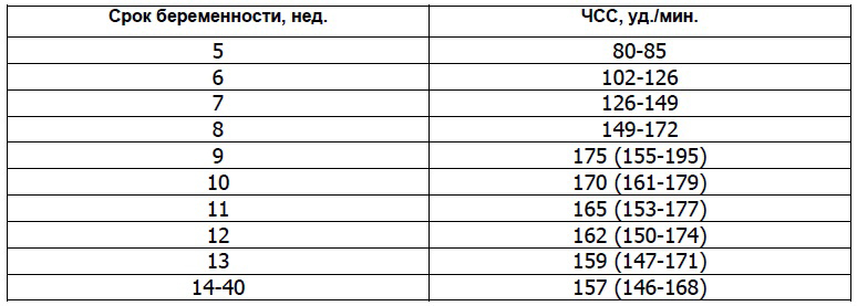
However, already in the first month of intrauterine life, the number of heartbeats of the baby will gradually increase (by about 3 beats per minute every day) and by the ninth week of pregnancy it will reach 175 beats per minute. Determine the fetal heart rate using ultrasound.
During the first ultrasound, specialists pay attention not only to the heart rate, but also to see how the baby's heart develops. To do this, use the so-called four-chamber cut , i.e. method of instrumental diagnosis of malformations of the heart.
It is important to emphasize that the deviation from the standards of such an indicator as heart rate indicates the presence malformations in the development of the heart . Therefore, doctors carefully study the structure on the cut atrial And fetal cardiac ventricles . If any abnormalities are found, the specialists refer the pregnant woman for additional studies, for example, echocardiography (ECG) with dopplerography.
Starting from the twentieth week, the gynecologist of the antenatal clinic will listen to the baby's heart with the power of a special tube at each scheduled visit to the pregnant woman. Such a procedure as auscultation of the heart not applied at earlier dates due to its inefficiency, tk. The doctor just can't hear the heartbeat.
However, as the baby develops, his heart will be heard more and more clearly each time. Auscultation helps the gynecologist determine the position of the fetus in the womb. For example, if the heart is better heard at the level of the mother's navel, then the child is in a transverse position, if the navel is to the left or lower, then the fetus is in cephalic presentation , and if above the navel, then in pelvic .
From the 32nd week of pregnancy, to control the heartbeat, use cardiotocography (abbreviated KTR ). When conducting the above types of examinations, a specialist can record in the fetus:
- bradycardia , i.e. abnormally low heart rate which is usually temporary. This deviation may be a symptom of the mother's autoimmune diseases, anemia, , as well as clamping the umbilical cord, when the unborn child does not receive enough oxygen. The cause of bradycardia can be congenital heart defects in order to exclude or confirm this diagnosis, a woman is necessarily sent for additional examinations;
- , i.e. high heart rate. Such a deviation is rarely recorded by specialists. However, if the heart rate is much higher than prescribed by the norms, then this indicates the mother or hypoxia , development intrauterine infections, anemia and genetic abnormalities at the fetus. In addition, medications a woman takes can affect heart rate.
In addition to the characteristics discussed above, when conducting the first screening ultrasound, specialists also analyze the data:
- about symmetry cerebral hemispheres fetus;
- about the size of the circumference of his head;
- about the distance from the occipital to the frontal bone;
- about the length of the bones of the shoulders, hips and forearms;
- about the structure of the heart;
- about the location and thickness of the chorion (placenta or "baby place");
- about the amount of water (amniotic fluid);
- about the state of the pharynx cervix mothers;
- about the number of vessels in the umbilical cord;
- about the absence or presence uterine hypertonicity .
As a result of ultrasound, in addition to the genetic abnormalities already discussed above ( monosomy or Shereshevsky-Turner syndrome, trisomy on chromosomes 13, 18 and 21 , namely Down, Patau and Edwards syndromes ) the following pathologies in development can be detected:
- neural tube , for example, spinal malformation (meningomyelocele and meningocele) or craniocerebral hernia (encephalocele) ;
- Cornet de Lange syndrome , an anomaly in which multiple malformations are fixed, entailing both physical abnormalities and mental retardation;
- triploidy , a genetic malformation in which a failure occurs in the chromosome set, as a rule, the fetus does not survive in the presence of such a pathology;
- omphalocele , embryonic or umbilical hernia, pathology of the anterior abdominal wall, in which some organs (liver, intestines, and others) develop in a hernial sac outside the abdominal cavity;
- Smith-Opitz syndrome , a genetic deviation that affects the processes, which subsequently leads to the development of many severe pathologies, for example, or mental retardation.
Biochemical screening of the 1st trimester
Let's talk in more detail about the second stage of a comprehensive screening examination of pregnant women. What it is biochemical screening of the 1st trimester, And what are the standards set for its main indicators? Actually, biochemical screening - is nothing but biochemical analysis blood of the expectant mother.
This study is carried out only after ultrasound. This is due to the fact that, thanks to an ultrasound examination, the doctor determines the exact gestational age, on which the standard values of the main indicators of blood biochemistry directly depend. So, remember that you need to go for biochemical screening only with the results of an ultrasound scan.
How to prepare for your first pregnancy screening
We talked about how they do it, and most importantly, when they do a screening ultrasound, now you should pay attention to preparing for the biochemical analysis. As in the case of any other blood test, this study must be prepared in advance.
If you want to get a reliable result of biochemical screening, you will have to follow the following recommendations exactly:
- blood for biochemical screening is taken strictly on an empty stomach, doctors do not even recommend drinking plain water, not to mention any food;
- a few days before the screening, you should change your usual diet and begin to follow a sparing diet, in which you can not eat too fatty and spicy foods (so as not to increase the level), as well as seafood, nuts, chocolate, citrus fruits and other allergenic foods, even if you have not previously had an allergic reaction to anything.
Strict adherence to these recommendations will provide a reliable result of biochemical screening. Believe me, it’s better to be patient for a while and give up your favorite treats so that you don’t worry about the results of the analysis later. After all, any deviation from the established norms, doctors will interpret as a pathology in the development of the baby.
Quite often, in various forums dedicated to pregnancy and childbirth, women talk about how the results of the first screening, expected with such excitement, turned out to be bad, and they were forced to do all the procedures again. Fortunately, in the end, the pregnant women received good news about the health of their babies, since the adjusted results showed the absence of any developmental abnormalities. 
The whole point was that expectant mothers were not properly prepared for screening, which ultimately led to inaccurate data.
Imagine how many nerves were spent and bitter tears were shed while women were waiting for new test results.
Such colossal stress does not pass without a trace for the health of any person, and even more so for a pregnant woman.
Biochemical screening of the 1st trimester, interpretation of the results
When conducting the first biochemical screening analysis, indicators such as free β-subunit of human chorionic gonadotropin (Further hCG ), as well as PAPP-A (plasma protein A associated with pregnancy) . Let's consider each of them in detail.
PAPP-A - what is it?
As mentioned above, PAPP-A - This is an indicator of a biochemical blood test of a pregnant woman, which helps specialists to establish at an early stage the presence of genetic pathologies in the development of the fetus. The full name of this quantity sounds like pregnancy-associated plasma protein A , which in literal translation into Russian means - pregnancy-associated plasma protein A .
It is protein (protein) A, produced during pregnancy by the placenta, that is responsible for the harmonious development of the unborn child. Therefore, an indicator such as the level of PAPP-A, calculated at 12 or 13 weeks during pregnancy, is considered a characteristic marker for determining genetic abnormalities.
It is mandatory to undergo an analysis to check the level of PAPP-A should:
- pregnant women over the age of 35;
- women who have previously given birth to children with genetic abnormalities;
- expectant mothers in whose family there are relatives with genetic abnormalities in development;
- women who have had diseases such as , or shortly before pregnancy;
- pregnant women who have had complications or spontaneous miscarriages in the past.
Normative values of such an indicator as PAPP-A depend on the gestational age. For example, the PAPP-A rate at 12 weeks is 0.79 to 4.76 mU/mL, and at 13 weeks is 1.03 to 6.01 mU/mL. In cases where, as a result of the test, this indicator deviates from the norm, the doctor prescribes additional studies.
If the analysis revealed a low level of PAPP-A, then this may indicate the presence chromosomal abnormalities in child development, for example, down syndrome, also it signals the risk of spontaneous miscarriage and regressive pregnancy . When this indicator is increased, this is most likely the result of the fact that the doctor could not calculate the correct gestational age.
That is why blood biochemistry is taken only after an ultrasound scan. However, high PAPP-A may also indicate the likelihood of developing genetic abnormalities in the development of the fetus. Therefore, in case of any deviation from the norm, the doctor will refer the woman for an additional examination.
Scientists gave this name to this hormone not by chance, because it is thanks to him that you can reliably find out about pregnancy already 6-8 days after fertilization has occurred. eggs. It is noteworthy that hCG starts to develop chorion already in the first hours of pregnancy.
Moreover, its level is growing rapidly and by the 11-12th week of pregnancy it exceeds the initial values by thousands of times. Then gradually loses its position, and its indicators remain unchanged (starting from the second trimester) until childbirth. All pregnancy test strips contain hCG.

If the level human chorionic gonadotropin increased, this may indicate:
- about the presence of the fetus down syndrome ;
- about multiple pregnancy ;
- about the development of the mother;
When the level of hCG is below the stipulated standards, it says:
- about a possible Edwards syndrome in the fetus;
- about risk miscarriage ;
- about placental insufficiency .
After the pregnant woman has undergone ultrasound and blood biochemistry, the specialist must decipher the results of the examination, as well as calculate the possible risks of developing genetic abnormalities or other pathologies using a special computer program PRISCA (Priska).
The screening summary form will contain the following information:
- about age risk anomalies in development (depending on the age of the pregnant woman, possible deviations change);
- about the values of biochemical parameters of a woman's blood test;
- about the risk of possible diseases;
- MoM coefficient .
In order to calculate as reliably as possible the possible risks of developing certain abnormalities in the fetus, experts calculate the so-called MoM (multiple of median) coefficient. To do this, all the obtained screening data is entered into a program that plots the deviation of each indicator of the analysis of a particular woman from the average norm established for most pregnant women.
MoM is considered normal if it does not go beyond the range of values from 0.5 to 2.5. At the second stage, this coefficient is adjusted taking into account age, race, presence of diseases (for example, diabetes ), bad habits (for example, smoking), the number of previous pregnancies, ECO and other important factors.
At the final stage, the specialist makes a final conclusion. Remember, only a doctor can correctly interpret screening results. In the video below, the doctor explains all the key points related to the first screening.
1st trimester screening price
The question of how much this study costs and where it is better to take it is of concern to many women. The thing is that not every state clinic can do such a specific examination for free. Based on the reviews left on the forums, many expectant mothers do not trust free medicine at all.
Therefore, you can often meet the question of where to do screening in Moscow or other cities. If we talk about private institutions, then in a fairly well-known and well-established INVITRO laboratory, biochemical screening can be done for 1600 rubles.
True, this cost does not include ultrasound, which the specialist will definitely ask to present before conducting a biochemical analysis. Therefore, you will have to separately undergo an ultrasound examination in another place, and then go to the laboratory for blood donation. And it must be done on the same day.
Second screening during pregnancy, when to do and what is included in the study
According to the recommendations of the World Health Organization (hereinafter referred to as WHO), every woman is required to undergo three screenings throughout the entire period of pregnancy. Although in our time, gynecologists refer all pregnant women to this examination, there are those who, for whatever reason, skip screening.
However, for some categories of women, such a study should be mandatory. This applies primarily to those who have previously given birth to children with genetic abnormalities or malformations. In addition, it is mandatory to undergo screening:
- women over the age of 35, since the risk of developing various pathologies in the fetus depends on the age of the mother;
- women who in the first trimester took drugs or other illegal drugs for pregnant women;
- women who have previously suffered two or more miscarriages;
- women who suffer from one of the following diseases that are inherited to the child - diabetes mellitus, diseases of the musculoskeletal system and the cardiovascular system, as well as oncopathology;
- women who are at risk of spontaneous miscarriage.
In addition, expectant mothers should definitely undergo screening if they or their spouses were exposed to radiation before conception, and also suffered immediately before or during pregnancy. bacterial and infectious diseases . As with the first screening, the second time, the expectant mother must also do an ultrasound and pass a biochemical blood test, which is often called a triple test.
Timing of the second screening during pregnancy
So, let's answer the question of how many weeks do the second screening
during pregnancy. As we have already determined, the first study is carried out in the early stages of pregnancy, namely in the period from 11 to 13 weeks of the first trimester. The next screening test is carried out during the so-called "golden" period of pregnancy, i.e. in the second trimester, which starts at 14 weeks and ends at 27 weeks. 
The second trimester is called golden because it is during this period of time that all the initial ailments associated with pregnancy ( nausea, weakness, and others) recede, and a woman can fully enjoy her new state, because she feels a powerful surge of strength.
A woman should visit her gynecologist every two weeks so that he can monitor the progress of the pregnancy.
The doctor gives recommendations to the future mother regarding her interesting situation, and also informs the woman about what examinations and for how long she should undergo. As a standard, a pregnant woman takes a urine test and a complete blood count before each visit to the gynecologist, and the second screening takes place from 16 to 20 weeks of pregnancy.
Ultrasound screening 2nd trimester - what is it?
During the second screening first, an ultrasound scan is performed to determine the exact gestational age, so that later specialists can correctly interpret the results of a biochemical blood test. On the ultrasound the doctor studies the development and size of the internal organs of the fetus: the length of the bones, the volume of the chest, head and abdomen, the development of the cerebellum, lungs, brain, spine, heart, bladder, intestines, stomach, eyes, nose, as well as the symmetry of the structure of the face.
In general, everything that is visualized with the help of an ultrasound examination is subjected to analysis. In addition to studying the main characteristics of the development of the baby, experts check:
- how the placenta is located;
- the thickness of the placenta and the degree of its maturity;
- the number of vessels in the umbilical cord;
- condition of the walls, appendages and cervix;
- quantity and quality of amniotic fluid.
Norms for ultrasound screening of the 2nd trimester of pregnancy:
Deciphering the triple test (biochemical blood test)
In the second trimester, experts pay special attention to three markers of genetic abnormalities, such as:
- chorionic gonadotropin - this is produced by the fetal chorion;
- alpha-fetoprotein ( Further AFP ) - this plasma protein (protein), initially produced yellow body, and then produced fetal liver and gastrointestinal tract ;
- free estriol ( further hormone E3 ) is a hormone produced in placenta , as well as fetal liver.
In some cases, they also study the level inhibin (hormone) produced follicles) . For each week of pregnancy, certain standards are established. It is considered optimal to conduct a triple test at 17 weeks of gestation.
When the level of hCG during the second screening is too high, this may indicate:
- about multiple pregnancy ;
- about diabetes at mother;
- about the risk of developing down syndrome if the other two indicators are below normal.
If hCG, on the contrary, is lowered, then this says:
- about risk Edwards syndrome ;
- about frozen pregnancy;
- about placental insufficiency .
When AFP levels are high, there is a risk of:
- anomalies in development kidney ;
- defects neural tube ;
- developmental disabilities abdominal wall ;
- damage brain ;
- oligohydramnios ;
- fetal death;
- spontaneous miscarriage;
- occurrence Rhesus conflict .
Decreased AFP can be a signal:
- Edwards syndrome ;
- diabetes mothers;
- low location placenta .
At a low level, the risk is high:
- development anemia in the fetus;
- adrenal and placental insufficiency;
- spontaneous miscarriage ;
- availability down syndrome ;
- development intrauterine infection ;
- delays in the physical development of the fetus.
It should be noted that at the level hormone E3 some drugs (for example,), as well as improper and unbalanced nutrition of the mother, affect. When E3 is elevated, doctors diagnose diseases kidney or multiple pregnancy, and also predict preterm birth, when the level of estriol rises sharply.
After the expectant mother goes through two stages of the screening examination, doctors analyze the information received using a special computer program and calculate the same MoM coefficient as in the first study. The conclusion will indicate the risks for a particular type of deviation.
Values are given as a fraction, such as 1:1500 (i.e. one in 1500 pregnancies). It is considered normal if the risk is less than 1:380. Then the conclusion will indicate that the risk is below the cut-off threshold. If the risk is higher than 1:380, then the woman will be referred for additional consultation with geneticists or offered to undergo invasive diagnostics.
It is worth noting that in cases where the biochemical analysis corresponded to the norms during the first screening (indicators were calculated HCG and PAPP-A ), then for the second and third time it is enough for a woman to do only an ultrasound.
The last screening examination of the expectant mother takes place in third trimester . Many people wonder what they look at at the third screening and when this study should be done.
As a rule, if a pregnant woman was not diagnosed with any abnormalities in the development of the fetus or during pregnancy at the first or second examination, then she only has to undergo an ultrasound examination, which will allow the specialist to draw final conclusions about the condition and development of the fetus, as well as his position in the womb. 
Determining the position of the fetus ( head or breech presentation ) is considered an important preparatory stage before childbirth.
In order for the delivery to be successful, and the woman to give birth on her own without surgical intervention, the child must be in the head presentation.
Otherwise, doctors plan a caesarean section.
The third screening includes procedures such as:
- ultrasound , which is passed by all pregnant women without exception;
- dopplerography is a technique that focuses mainly on the condition of the vessels placenta ;
- cardiotocography - a study that allows you to more accurately determine the heart rate of a child in the womb;
- blood biochemistry , during which attention is focused on such markers of genetic and other abnormalities as the level hCG, α-fetoprotein and PAPP-A .
Timing of the third screening during pregnancy
It is worth noting that only the doctor decides how many weeks 3 screening a woman should undergo, based on the individual characteristics of this particular pregnancy. However, it is considered optimal when the expectant mother undergoes a planned ultrasound at 32 weeks, and then immediately passes a biochemical blood test (if indicated), and also undergoes other necessary procedures.
However, for medical reasons, dopplerography or KTG fetus can be from the 28th week of pregnancy. third trimester starts at 28 weeks and ends with childbirth at 40-43 weeks. The last screening ultrasound is usually prescribed at 32-34 weeks.
Deciphering ultrasound
At what time the third screening ultrasound passes a pregnant woman, we found out, now let's talk in more detail about the decoding of the study. When performing an ultrasound in the third trimester, the doctor pays special attention to:
- for the development and construction of cardio-vascular system a child to exclude possible developmental pathologies, for example,;
- for proper development brain , organs of the abdominal cavity, spine and genitourinary system;
- to those in the cranial cavity vein of Galen , which plays an important role in the proper functioning of the brain to eliminate aneurysm ;
- on the structure and development of the face of the child.
In addition, ultrasound allows a specialist to assess the condition amniotic fluid, appendages and uterus mothers, as well as check and placenta thickness . In order to exclude hypoxia and pathologies in the development of the nervous and cardiovascular systems , as well as to identify the features of blood flow in uterine vessels and the child, as well as in the umbilical cord, carry out dopplerography .
As a rule, this procedure is carried out only according to indications simultaneously with ultrasound. In order to exclude fetal hypoxia and define heart rate, carry out KTG . This type of research focuses solely on the functioning of the baby's heart, so cardiotocography prescribed in cases where the doctor has concerns about the condition cardiovascular child systems.
Ultrasound in the third trimester of pregnancy allows you to determine not only the presentation of the child, but also the maturity of his lungs, on which the readiness for birth depends. In some cases, to save the life of the child and the mother, hospitalization may be required for the purpose of early delivery.
| Indicator | The average rate for 32-34 weeks of pregnancy |
| Placenta thickness | from 25 to 43 mm |
| Amniotic (amniotic) index | 80-280 mm |
| Degree of placental maturity | 1-2 degree of maturation |
| Uterine tone | missing |
| uterine pharynx | closed, length not less than 3 cm |
| Fetal growth | average 45 cm |
| Fetal weight | on average 2 kg |
| Girth of the abdomen of the fetus | 266- 285 mm |
| BDP | 85-89 mm |
| Fetal thigh length | 62-66 mm |
| fetal chest circumference | 309-323 mm |
| Fetal forearm size | 46-55 mm |
| Fetal leg bone size | 52-57 mm |
| Fetal shoulder length | 55-59 mm |
According to the results of a biochemical blood test MoM factor should not deviate from the range from 0.5 to 2.5. The risk value for all possible deviations must correspond to 1:380.
Education: Graduated from Vitebsk State Medical University with a degree in Surgery. At the university, he headed the Council of the Student Scientific Society. Advanced training in 2010 - in the specialty "Oncology" and in 2011 - in the specialty "Mammology, visual forms of oncology".
Work experience: Work in the general medical network for 3 years as a surgeon (Vitebsk Emergency Hospital, Liozno Central District Hospital) and part-time as a district oncologist and traumatologist. Work as a pharmaceutical representative for a year in the Rubicon company.
He presented 3 rationalization proposals on the topic “Optimization of antibiotic therapy depending on the species composition of microflora”, 2 works won prizes in the republican competition-review of student scientific works (categories 1 and 3).
First screening during pregnancy- a set of examinations, the purpose of which is to identify the risk of congenital pathologies of the fetus. The diagnostic test includes an ultrasound and a blood test from a vein. The optimal timing for its implementation coincides with the end of the first trimester of pregnancy.
Prenatal screening shows the risk of pathology in the unborn child, but it cannot say with absolute certainty about its presence or absence. Deviations from the norm in its results are an indication for other diagnostic studies, during which specialists will make a final diagnosis.
With the help of screening of the 1st trimester, the expectant mother can find out about a severe congenital anomaly of the fetus and decide on time to terminate the pregnancy.
Deadlines for the study
Screening in the first trimester of pregnancy can only be carried out from the first day of week 11 to the sixth day of week 13. It is not advisable to conduct studies at an earlier date, since the structures examined on ultrasound are still too small for observation, and the hormones of the biochemical test have not reached the desired concentration.An ultrasound examination at a later date does not make sense. This phenomenon is due to the fact that after the lymphatic system begins to develop in the fetal body, the fluid of which interferes with the consideration of anatomical structures.
The optimal timing for the first screening corresponds to 11-12 weeks of pregnancy. At this time, biochemical tests have the smallest error, and with the help of ultrasound equipment, the anatomical structures of the unborn child are clearly visible. Also, if malformations are detected, a woman can have time to make a medical termination of pregnancy in a less traumatic way.
Indications for ultrasound and blood tests
According to the order of the Ministry of Health of the Russian Federation, screening of the 1st trimester is indicated for all pregnant women. It helps to identify malformations in the early stages of gestation and is absolutely safe for the body of the fetus and expectant mother. Every pregnant woman has the right to decide whether she will undergo prenatal screening.Mandatory first screening is recommended for women in one of the risk groups:
- age over 40;
- history of spontaneous abortion;
- intrauterine fetal death during previous pregnancies;
- the birth of a child with a history of chromosomal abnormalities;
- transferred infectious and inflammatory disease during the period of gestation;
- the use of drugs that have a teratogenic (causing congenital anomalies) effect on the fetus during the gestation period;
- alcohol abuse or drug use during the gestation period;
- burdened hereditary history (the presence of congenital anomalies in close relatives);
- having a blood relationship with the father.
Goals of the first screening
The main goal of screening in the first trimester is to identify the degree of risk of congenital pathology of the fetus. With the help of ultrasound equipment, a specialist examines the "markers" of genetic diseases. Their presence indicates a high probability of chromosomal abnormalities.Biochemical screening evaluates the amount of hormones produced by the placenta. It is called "double" because it consists of counting hCG and PAPP-A. Their deviation from normal values indicates a high probability of congenital diseases.
With the help of the studies described, specialists can establish an increased risk of certain chromosomal abnormalities - Edwards, Patau, Down, de Lange, etc. syndrome. Their danger lies in the fact that many children die in the first months and years after birth, because their organs have an atypical structure and cannot fully perform their functions. But if the child manages to survive, his mental and physical development will lag far behind his peers.
Attention! Both examinations of the first screening cannot say for sure whether the fetus has a chromosomal anomaly, these tests only reflect the risk of its presence, therefore, with poor results, expectant mothers should not fall into despair - quite often the alarm is in vain.
High risk at the first screening is assessed by the width of the neck crease. Normally, it should not exceed 0.3 centimeters. The greater the deviation from this value, the higher the likelihood of a chromosomal abnormality.
Another marker of Down syndrome is the structure of the nasal bone. The high risk of a chromosomal abnormality is indicated by its absence at any stage of pregnancy. After 12 weeks, doctors measure the length of the nasal bone, normally it should exceed 3 mm. The shorter dimensions of this anatomical structure are a marker of chromosomal pathology.
In addition to chromosomal pathologies, screening helps in the diagnosis of neural tube defects. This group of diseases is characterized by abnormal laying of the brain or spinal cord, which is rarely compatible with life. Also, ultrasound examination can reveal anomalies of other organs - the absence of limbs, the laying of the heart outside the chest cavity, a hernial protrusion of the anterior abdominal wall, etc.
Preparing for the first screening
Ultrasound screening can be done in two ways. The first of them - transvaginal - when the sensor is inserted into the vagina. With this type of ultrasound, a woman does not require special preparation.The second way to conduct an ultrasound is transabdominal - when the sensor is located on the surface of the abdomen. In this case, for reliable results, the bladder must be filled, so preparation for the first screening includes taking one liter of fluid one hour before the study.
To pass the second stage of screening in the last weeks of the 1st trimester, the expectant mother should donate blood from a vein. The sampling of material for biochemical research is carried out in the morning. For reliable results a woman should not eat breakfast before the analysis. On the day of blood donation, only one glass of pure non-carbonated water is allowed.
Three days before taking blood for a biochemical analysis, a woman is recommended to exclude allergens from the diet. These include seafood, peanuts, milk, fish, chocolate. It is also undesirable to eat fried, salty, smoked foods - this food can distort the results of the study.
To obtain reliable results, a woman should try to relax, since the increased activity of the nervous system can affect the production of placental hormones. Before research, you should sleep well and rest. Also many experts recommend to exclude sexual activity three days before the proposed screening.

Features of the
Usually screening in the first trimester is carried out in three stages. The first of them - preparatory - includes a survey of complaints and an examination by an obstetrician-gynecologist. The specialist collects the life and hereditary history of the woman, calculates the date of conception. For the reliability of the screening results, the doctor must know the exact age of the patient, her chronic diseases, endocrine pathologies, and the presence of IVF.Without fail, the doctor finds out if there are hereditary diseases in the family. The specialist will also learn about the course of previous pregnancies - the presence of miscarriages, the birth of a child with congenital anomalies, etc. Expectant mothers with a burdened hereditary history are shown genetic screening.
After consulting a doctor, a woman is given a referral for ultrasound and biochemical analysis. In different clinics, the sequence of their implementation is different.
Sometimes both tests are done on the same day. Most often this happens if the ultrasound is done transvaginally, because a large amount of water must be consumed before the transabdominal ultrasound. This can skew the results of the first screening for placental hormones.
Sometimes the study is carried out in two stages. On the first day, a woman undergoes an ultrasound scan, which calculates the date of conception. The values obtained are necessary to decipher the results of the biochemical test, since the amount of hormones changes every day.
Less commonly, women first donate blood for a biochemical test. With the results obtained, the expectant mother goes to an ultrasound scan, where the gestation period is calculated again.

ultrasound screening
Usually, a fetal ultrasound scan does not exceed 30 minutes. If the study is performed transvaginally, a disposable condom is put on the sensor, then it is inserted into the vagina. When performed correctly, a pregnant woman should not experience discomfort.With transabdominal ultrasound, the front wall of the abdomen of the expectant mother is lubricated with a special gel. The sensor of the device will slide on it, the image is shown on the monitor. Such a study also does not cause discomfort in a pregnant woman.
Many women ask to determine the sex of the child at the first screening. Some doctors may try to look at the pelvic area of the fetus. However, for accurate results, the baby must be facing the anterior uterine wall.
The longer the gestation period, the higher the probability of correctly determining the sex of the child. At week 11, the number of successful attempts does not exceed 50%. At the end of the 13th week, in 80% of cases, the doctor can determine the sex of the baby.
To perform ultrasound screening, certain conditions must be met. The first of them is the length of the fetus from the coccyx to the crown of at least 4.5 centimeters. Secondly, the unborn child must take the desired position in the uterine cavity. To do this, the doctor may ask the woman to move or cough.
On an ultrasound examination, the doctor evaluates the following main parameters:
- KTP - the length of the fetus from the end of the spine to the crown;
- skull circumference;
- BPR - the length of the space between the parietal tubercles;
- TVP - thickness of the collar space (neck fold);
- pulse;
- the length of the bones of the limbs;
- presence, position, structure of internal organs;
- anatomical structure of the placenta;
- the presence and structure of the nasal bone.
Gestational age
Nasal bone, mm
Pulse, beats/min
If there are indications or at the request of a pregnant woman, the doctor can conduct an additional study using a Doppler transducer. This test shows the state of blood exchange between the vessels of the uterus and placenta, pathologies indicate oxygen starvation of the fetus. Doppler ultrasound also allows you to see the number of vessels in the umbilical cord - normally there should be two arteries and one vein.
Biochemical screening
To assess the amount of hormones produced by the placenta, specialists take blood from a vein. This procedure is almost painless, in the presence of fear, you should not look at the syringe. First, the laboratory assistant puts a tourniquet on her shoulder, then the woman needs to clench her fist several times. After the described manipulations, the specialist inserts a needle into the vein and takes a few milliliters of blood.The screening evaluates the amount of human chorionic gonadotropin. This hormone is produced by the placenta, its amount in the blood increases up to 11 weeks, then it decreases slightly. Many chromosomal and placental abnormalities are accompanied by a change in the amount of hCG.