Late gestosis (toxicosis) in pregnant women is a pathological condition when all systems of a woman’s body cannot cope with the fetal needs for nutrients and oxygen. This condition begins after the twentieth week of pregnancy and may last several weeks after delivery. The main symptoms of gestosis are: edema, high blood pressure and proteinuria. Late toxicosis in pregnant women can manifest itself as one symptom, but sometimes the symptoms are combined. In this regard, four forms of gestosis are distinguished:
- dropsy;
- nephropathy (1,2,3 degrees);
- preeclampsia;
- eclampsia.
These forms of late toxicosis are typical and are considered by doctors as different stages of the same process.
Dropsy
This is the earliest stage of gestosis, which is characterized by the manifestation of only one symptom - edema. They are usually widespread: swelling of the lower and upper extremities, the anterior abdominal wall, and puffiness or swelling of the face may also appear. With dropsy, the general condition of pregnant women is not disturbed; only in cases where the swelling is too pronounced, women may experience a slight feeling of heaviness. It is quite difficult to diagnose edema as the first stage of gestosis, since they also appear during a normally ongoing pregnancy. You need to weigh yourself regularly in order to notice rapid weight gain (more than 0.5 kg per week) in time, since swelling can also appear inside the body.
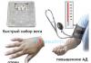
In 20-24% of cases, dropsy develops into nephropathy, a stage characterized by a combination of several symptoms: edema, hypertension and proteinuria. The main symptom of nephropathy is high blood pressure (), which can sometimes reach 200/150 mmHg. Art., and sometimes higher. The degree of this stage of gestosis is determined according to the Savelyeva scale, which characterizes the condition of pregnant women during examination. There are three main degrees of nephropathy.
Preeclampsia 1st degree
Edema appears slightly, mainly in the lower extremities. Mild hypertension appears - about 150/90 mm Hg. Art., protein appears in the urine - up to 1.0 g/l.
Gestosis 2 degrees
Edema spreads to the lower extremities and abdominal cavity. Blood pressure rises to 170/100 mmHg. Art., protein in urine – 1.0-3.0 g/l.
Gestosis 3 degrees
Abdomen, face. Blood pressure – more than 170/100 mm Hg. Art., protein in urine – more than 3.0 g/l. Hemorrhages and degenerative changes may appear in the fundus.
Preeclampsia
Preeclampsia is a more severe form of late toxicosis. To the three symptoms of nephropathy (edema, hypertension, proteinuria), others are added: dizziness, headache, insomnia, apathy, blurred vision (small “spots”, “veil” before the eyes, sometimes complete loss of vision), lethargy, tinnitus and sensation heaviness in the back of the head or forehead. There are also signs of digestive tract and liver disorders: nausea, abdominal pain, vomiting. In this condition, a pregnant woman can experience eclampsia (convulsive seizure) from any irritant (bright light, loud sound, pain).
Eclampsia
This stage of gestosis is the most dangerous for the pregnant woman and the fetus, but its cases are rare. Whole body seizures are accompanied by severe fluctuations in blood pressure, which can lead to cerebral hemorrhage (stroke), placental abruption, and fetal death. Also, a pregnant woman may experience acute respiratory failure (difficulty breathing, agitation, shortness of breath). Eclampsia leads to loss of consciousness and occurs in four main stages:
Stage 1 – twitching of the facial muscles is observed for 20-30 seconds.
Stage 2 – tonic convulsions are observed for 2-30 seconds (prolonged muscle contraction, as a result of which the limbs “freeze” in the position of extension or flexion, the head is brought to the chest or thrown back, the patient’s body is stretched). This stage can lead to biting the tongue and even stopping breathing.
Stage 3 – for about two minutes, the pregnant woman experiences clonic convulsions (repeated contraction of the flexor and extensor muscles), cyanosis develops (blue discoloration of the skin), breathing is impaired, and foamy saliva with blood appears.
Stage 4 is the final stage, which begins with a deep breath. The pregnant woman may not yet regain consciousness.
Preeclampsia in pregnant women can occur slowly, with mild symptoms, or it can progress very quickly and develop into eclampsia within a few days. In both cases, fetal hypoxia occurs.
© Use of site materials only in agreement with the administration.
Any woman who has given birth is familiar with some features of the course of pregnancy and the main stages of monitoring it: regular visits to a medical facility, testing, ultrasound examinations, weighing. Some people are surprised by the need for weight control. Like, why should medical workers care about the future figure of a pregnant woman? Any diagnostic procedure has a meaning and is conditioned by something.
How many kilograms should a woman's weight increase during pregnancy? Many will answer the question correctly - about 10 kg. What if it’s 20–25? Such an increase “speaks” of hidden (and not only) edema. And swelling is gestosis. For women, this disease is more commonly known as late toxicosis.
Edema is one of the diagnostic signs of gestosis, but the pathology is not limited to them. Preeclampsia is easier in healthy women. In this case it is called “pure”. This type of disease occurs in 30% of pregnant women. If it develops against the background of existing diseases (hypertension, diabetes, gastrointestinal ailments, kidney diseases), then in this case they speak of “combined” gestosis. It is clear that the latter form is more difficult.

The first signs of possible gestosis
This pathology concerns only pregnant women - after childbirth, gestosis goes away. However, gestosis is classified as a dangerous disease. Its cunning lies in its complications. A quarter of female deaths during pregnancy are due to gestosis. The fetus dies 3-4 times more often than during an uncomplicated pregnancy.
The main cause of gestosis is a dysregulation of blood vessels, resulting in their spasm. Microvessels are mainly affected.
As for the pathogenesis of gestosis: many scientists see its connection with immune factors. Fetal antigens influence maternal antibody production. In turn, antibodies cause the formation of excess immune complexes, which have a negative effect on the pregnant woman’s body.
How does gestosis manifest?
The disease is often called OPG-preeclampsia. OPG – the first letters of the terms: edema, proteinuria, . These are the main signs of pathology. The entire complex is not always noted. Mild gestosis can occur with one or two of the listed symptoms.

OPG complex of symptoms
Swelling is more common in pregnant women. A woman drinks a lot of fluid, which cannot completely leave the body and lingers in the interstitial space. Only the lower extremities may swell, but in more severe forms, the entire body may swell. Swelling is not always noticeable. Sometimes we are talking about a hidden form. It is detected by weighing. A weight gain of more than 0.5 kg per week indicates an emerging problem. Monitoring of fluid intake and amount of urine excreted is prescribed. If, during normal drinking conditions, less than 0.8 liters of urine is excreted, preeclampsia can be suspected.
Hypertension develops against the background of fluid retention. Blood pressure is monitored at every doctor's visit. Pressure during gestosis exceeds the norm by 15–20%. What pressure is considered normal? Usually it is 120/80. However, if a woman had symptoms at the beginning of pregnancy, then even the generally accepted norm for her may already be a signal for further examination.
Proteinuria refers to the excretion of protein in the urine. This sign indicates a violation of renal function. Therefore, it is important not to skip a diagnostic procedure such as a urine test. After 20 weeks of pregnancy, urine is examined weekly.
If a woman has two of the three signs of this disease, then treatment at home is ineffective - it is better to hospitalize the patient.
Other symptoms include headache, vomiting, nausea, and heaviness in the head. In the most difficult cases - changes in consciousness and convulsions.
The manifestation of pathology in pregnant women is more likely:
- Expecting their first child;
- Having genital tract infections: chlamydia, mycoplasmosis, ureaplasmosis;
- Suffering from chronic diseases: hypertension, diabetes, kidney disease, excess weight and others;
- Expecting twins.
Classification of gestosis
One of the classifications of pathology is divided into types:
- Early gestosis;
- Late gestosis.
The disease becomes more severe at the end of pregnancy.
Depending on the signs and form, the disease can be divided into the following degrees of severity:
1st degree
Dropsy of pregnancy is classified as gestosis of the 1st degree. This stage is characterized only by edema of varying severity. Usually they are less pronounced in the morning, and in the evening the condition worsens.
2nd degree
With grade 2 gestosis, all three symptoms of OPG are observed. In diagnosing hypertension, the most important indicators are diastolic pressure. The fact is that it is directly related to placental blood flow: the higher the diastolic pressure, the less oxygen the child receives. It is noteworthy that it is not so much the increase in pressure that is dangerous as its abrupt changes. This stage is especially difficult for pregnant women with concomitant diseases.
Complications develop:
- Placental abruption;
- Bleeding;
- Premature birth.
The main danger is that with complicated gestosis, the fetus is at risk of death.
Nephropathy is diagnosed simply by urine analysis. If things go wrong, it is important to monitor the condition of the fundus. Changes may indicate.
Stage 3, preeclampsia
As the condition worsens, stage 3 of gestosis develops. Pain and heaviness in the head indicate the onset of preeclampsia. Possible blurred vision, vomiting, and pain in the liver area. Memory deterioration, apathy, insomnia, irritability and other signs of changes in blood circulation in the brain are possible. Edema has a damaging effect on the liver, as evidenced by pain on the right side. There are even hemorrhages in this organ. “Floaters” and “veils” before the eyes may indicate problems with the retina.

Main signs of preeclampsia:
- The amount of urine decreases to 0.4 liters or less;
- Blood pressure – 160/110 or more;
- Protein in urine;
- Blood clotting disorder;
- Changes in liver function;
- Nausea, vomiting;
- Symptoms of brain and visual disorders.
Eclampsia
An even more severe degree of gestosis is eclampsia. In addition to all of the above symptoms, convulsions are present. Typically, attacks are caused by external stimuli: loud sound, bright light, stress, pain. The attack does not last long - about 2 minutes. The danger of this condition is cerebral edema and death. Despite the similarities between gestational seizures and epileptic seizures, they have a number of differences. In epilepsy, urine tests are normal, there is no hypertension, and a characteristic epileptic aura is noted before a seizure.
HELLP syndrome
One of the dangerous forms is called HELLP syndrome. Its signs include bloody vomiting, jaundice, severe coma, and liver failure. Usually observed in women who have given birth frequently. May occur even after childbirth(unlike other forms of gestosis). About 80% of women and the same number of unborn children die from this type of pathology.
The most rare forms of gestosis include:
- Eczema;
- Dermatoses;
- Bronchial asthma;
- Pregnancy itch.
Some researchers suggest that all these forms are exacerbations of pre-existing diseases in women.
With different frequencies, pregnant women may suffer from other types of gestosis:
- Osteomalacia. Otherwise - softening of the bones. A pronounced form is rare. More often it manifests itself in tooth decay, bone pain, changes in gait, and neuralgia. The reason for this condition lies in the lack of microelements - especially calcium - and vitamins.
- Ptyalism (salivation). It is often accompanied by vomiting. With excessive saliva production, the body becomes dehydrated, speech is impaired, and the skin and mucous membranes are irritated.
- Hepatosis. Accompanied by jaundice. It is necessary to differentiate with hepatitis. Therefore, a thorough diagnosis is carried out, and the woman is temporarily isolated from others.
- Liver atrophy. If such a complication occurs during early gestosis and cannot be treated, then it is recommended to terminate the pregnancy.
- HELLP syndrome is considered a truly rare form. Still, for most women, pregnancy ends happily - with the birth of a healthy baby.
Complications of gestosis
Mild gestosis can be almost invisible. Why get examined, let alone be hospitalized, if you feel good and don’t hurt anything! But I would like to emphasize that The main danger of the disease is its consequences, such as:
- Pulmonary edema;
- Hemorrhage;
- Pathology of the cardiovascular system;
- Placental abruption;
- Kidney diseases;
- Delay in child development;
- HELLP syndrome;
- Early birth;
- Liver diseases;
- Fetal hypoxia;
- Brain swelling;
- Problems with the retina of the eye;
- Stroke;
- Death of a child;
- Death of a pregnant woman.
Important! The development of dangerous stages of gestosis and their consequences can be prevented by timely diagnosis and correctly prescribed treatment.
Diagnostics
Every woman undergoes regular medical examination during pregnancy; if alarming symptoms appear, such examination is carried out unscheduled, and diagnostic procedures are added.
Required studies include:
- Weighing. In the second part of pregnancy, weight gain should not exceed 350 g per week. If a woman has gained half a kilogram or more, then additional examinations need to be carried out.
- The need to control fluid intake. For pregnant women, the rule “2 liters or more of water per day” is not suitable. And when pronounced edema appears, its amount should not be more than 1 liter. It is also necessary to control the volume of urine excreted.
- Carrying out a blood test. The number of platelets and red blood cells is determined. Particularly important is the indicator of platelet content and coagulation. In addition to the general one, a biochemical analysis is carried out.
- Blood pressure control, and on each arm. The presence of gestosis can be indicated by a large difference in the indicators on the left and right hands.
- Analysis of urine. It is necessary to monitor the presence of protein in the urine.
- Ultrasound of the fetus with. With the help of this study, the degree of fetal development and malnutrition is revealed.
- Dentist examination.
- Fundus examination. If the vessels of the fundus are changed, this may indicate problems with the vessels of the brain.

A woman should not ignore medical examinations. This is especially true for mature mothers (after 35 years) and those who are giving birth to their first child. Also, pregnant women with chronic and infectious diseases should be attentive to their health.
Successful diagnosis is the key to a properly structured treatment strategy.
Important! Not a single symptom should escape the attention of a pregnant woman. She must immediately report her suspicions to her doctor.
How to treat gestosis?
Let's say right away that Preeclampsia cannot be completely cured. It goes away with pregnancy. However, it is possible to prevent its development into more severe forms.
Main areas of treatment:
- It is necessary to create a protective treatment regime. A woman should avoid strong emotional stress, be calm and balanced. Bright light, noise, heavy physical activity that does not correspond to her condition are harmful. If gestosis is mild, then remedies such as motherwort and valerian are prescribed, and in more severe forms, they resort to individual selection of sedatives taking into account pregnancy.
- To restore the function of the pregnant woman’s body, as well as to prevent fetal hypoxia, appropriate medications are prescribed. These are medications that have a sedative, hypotensive, antispasmodic, and diuretic effect. Such drugs should improve placental blood flow, preventing fetal hypoxia. In case of exacerbation of existing chronic diseases, appropriate treatment is prescribed aimed at relieving symptoms.
- The birth canal must be prepared so that delivery during gestosis can be carried out in a timely and careful manner. The timing of delivery is determined by the condition of the pregnant woman. For example, a severe form of gestosis requires delivery no later than three days after the condition worsens. If eclampsia develops, delivery should be immediate. Childbirth is safest for the health of the baby at 38 weeks of pregnancy and later, since by this time all the vital systems of the fetus have time to form. Give birth naturally or use a caesarean section? The choice depends on the condition of the fetus and the birth canal of the pregnant woman. In case of severe gestosis, when urgent delivery is necessary, a caesarean section is performed. If childbirth occurs naturally, anesthesia is recommended. And not so much for pain relief, but to improve placental and renal circulation, as well as a slight decrease in pressure. In the case of mild gestosis, treatment is prescribed, and childbirth occurs on time naturally.
In moderate and mild forms of gestosis, women are recommended to be hospitalized in a hospital. Severe gestosis may require resuscitation. The main research procedures carried out in the hospital:
- Conducting a general urine test, as well as a Zimnitsky test.
- Study of the condition of the unborn child (Doppler, ultrasound, cardiotocography).
- Coagulogram and other blood tests.
Infusion therapy is used as treatment, the task of which is to remove tissue fluid, as well as replenish its deficiency in the vessels. For hypertension, individual selection of medications is carried out.
Treatment lasts from several hours to several weeks. It all depends on the severity of the condition. The more dangerous it is, the less time a woman has. Delivery is the main outcome of any treatment. Therefore, in the most severe cases, an immediate caesarean section is performed.
Principles of hexose prevention
The main task of the patient and medical workers is timely detection of the disease and initiation of early treatment. Therefore, it is difficult to avoid gestosis without active prevention.
 Avoid obesity. During pregnancy, women sometimes gain a lot of weight. Why is this happening? There are many reasons. Firstly, hormonal changes can cause an increase in appetite. Secondly, a woman begins to allow herself to eat everything, citing the fact that her figure is already deteriorating, it won’t get any worse. Thirdly, some women are sure that you need to eat for two. If she eats as before, the baby will not get many nutrients. Unfortunately, the effect of such eating behavior is sad - obesity and gestosis.
The diet for gestosis is very simple. The fetus needs protein (the cells of the unborn child’s body will be built from it), which means that a pregnant woman’s diet should include dairy products, lean meat, eggs, and fish. An increased protein content in food is also necessary because it migrates from the body.
You need vitamins, minerals, fiber. And they are most abundant in vegetables and fruits. Fiber is especially important: with a minimum of calories, it perfectly satisfies hunger. This diet is also beneficial for the gastrointestinal tract - there will be no constipation or complications such as those that often bother pregnant women. It’s good to forget about the existence of flour and sweet foods. Apart from excess weight, they will give nothing to either mother or child.
The maximum weight gain during the entire pregnancy should be no more than 12 kg. Women with initial underweight may gain a little more. Conversely, plump mothers are allowed to gain a maximum of 10 kg.
Proper drinking regime is very important. Despite the threat of edema, you should not severely limit yourself in water. It is recommended to consume 1 to 1.5 liters of fluid per day, this also includes fruits, soups and other foods. But you cannot retain this water with salt. No matter how much a pregnant woman would like to eat a pickled cucumber or a piece of herring, there is no need to do this. To remove excess fluid, as well as improve renal blood circulation, it is useful to drink a decoction of bearberry, rose hips, cranberry juice, kidney tea (by agreement with your doctor!). For the same purposes, the doctor may prescribe special medications: cystone, canephron, etc.
And one more, and perhaps the most important principle of prevention - active lifestyle. Pregnancy is not a disease. Therefore, a pregnant woman, like any other woman, should walk, swim, do yoga for pregnant women, Pilates, and not forget about special gymnastics. The main thing is not to overdo it. It is necessary to listen to your condition and stop exercising at the slightest suspicion of its deterioration. For your peace of mind, it is better to consult a doctor once again. Physical activity should not harm a woman and her unborn child. The doctor may recommend special exercises to help get rid of certain manifestations of the pathology.
Undiagnosed and untreated gestosis is dangerous. Only careful attention to yourself will allow a woman to give birth to a healthy child and maintain her own health.
Video: gestosis in the cycle “Pregnancy week by week”
- The condition is the main danger of the second half of pregnancy and poses a serious threat to the life of the expectant mother.
What it is? Preeclampsia during pregnancy is a pathological condition of the body in which the functions of vital organs are disrupted and which is extremely difficult to control if it has entered an advanced stage.
The disease occurs mainly in the third trimester and has another name - late toxicosis. However, it differs from the classic ailment in the form of nausea and vomiting in that it entails dysfunction of the cardiovascular and endocrine systems, damage to the central nervous system as a result of spasm of blood vessels.
The prevalence reaches 30%; the situation is further complicated by the fact that gestosis in the first half of pregnancy is very difficult to detect in the initial stages of development. For example, late toxicosis, which began at 20 weeks, is detected only by 27–28 weeks.
What is the danger of gestosis?
Until now, despite the development of medicine, gestosis remains one of the main causes of maternal and infant mortality in the prenatal and postpartum period. It does not kill instantly, but contributes to the rapid decline of the body over several days.
The patient may lose vision, the ability to move independently, and one by one important organs stop functioning: liver, kidneys, heart, brain. The more serious the stage of gestosis, the less chance doctors have to save the patient and (or) her child.
Only close attention to the deterioration of your health and timely examination will help identify late toxicosis in the early stages of its development and avoid the fatal risk.
Causes of gestosis
Scientists have not yet reliably found out what exactly is the cause of the development of late gestosis. There are only some assumptions about this:
- Pathological changes in the central nervous system. The relationship between the cerebral cortex and subcortical structures is disrupted, which leads to pathology. The trigger is the psychological stress that a woman may have been exposed to while carrying a child.
- Immune disorders, in particular, failures in the recognition of maternal tissue and fetal tissue. This process involves special T cells, which are regulators of the immune response.
- Disruptions in the endocrine system. Pregnancy involves dramatic changes in hormonal status, which can result in disruptions in the functioning of a woman’s entire body.
- Lack of folic acid. This provokes an increase in the level of non-proteinogenic amino acids, which are extremely toxic to the body.
Preeclampsia, a complication of pregnancy, involves spasm of all blood vessels - this is what causes the failure of vital organs.
Symptoms of gestosis during pregnancy by stage

There are several classifications of late gestosis, but doctors in Russia distinguish 4 main stages in the development of the disease, each of which is characterized by certain clinical manifestations.
Dropsy
It is characterized by insufficient removal of fluid from the body, resulting in swelling. This stage is divided into 4 stages, which are characterized by the ascending direction of localization of edema:
- The feet swell, and there is slight swelling of the legs.
- The legs swell completely, the lower third of the abdomen swells.
- The swelling rises higher and affects the face, in addition to the legs and body.
- Edema affects the entire body and is observed in internal organs.
Characteristic signs of edema
- When you press your finger on the surface of the skin, a dent remains. The longer it takes to disappear, the more severe the swelling becomes.
- There is tingling and numbness in the swollen limb.
- Severe swelling causes a feeling of fatigue in a pregnant woman.
These are the earliest symptoms of gestosis during pregnancy - if doctors prescribe the woman the necessary therapy, then late toxicosis will not develop further.
Nephropathy
If no therapeutic measures were taken when edema appeared, then the disease progresses and enters the stage of nephropathy. In addition to fluid retention, hypertension appears in the body, and urine tests indicate an increase in protein.
All these symptoms of gestosis will be noticeable to the doctor if the patient visits the antenatal clinic at least once every 2 weeks and regularly passes the necessary tests. Nephropathy has several degrees, which have certain symptoms:
- I degree - the pressure does not exceed 150/90, and the distance from the upper to the lower limit should be normal. A urine test reveals protein of no more than 1 g/l. There is swelling of the lower extremities.
- II degree - pressure does not exceed 170/100, protein in the urine increases and begins to reach 3 g/l. Edema spreads not only to the lower extremities, but also to the lower third of the abdominal wall.
- III degree – pressure is above 170/110, protein in the urine exceeds 3 g/l, swelling spreads throughout the body, swelling of the internal organs is detected.
Nephropathy, especially its severe degree, cannot go unnoticed, and the pregnant woman will be forced to go to the hospital due to the deterioration of her condition.
Preeclampsia

In some cases, stage III nephropathy, despite treatment, develops into preeclampsia. The main difference between this condition and nephropathy is that the pregnant woman has a circulatory disorder in the brain.
There is a real threat to the life of the mother and fetus, which requires immediate hospitalization. Among the signs of severe gestosis during pregnancy are the following:
- Confusion
- Headache
- Loss of vision and/or hearing
- Feeling of heaviness in the back of the head
- Manifestations of sclerosis
- Hemorrhages in the walls of vital organs
- Vomit
If a woman in this condition is left without medical care, she will die. Preeclampsia involves placing the patient in an intensive care unit, since her well-being must be monitored around the clock.
Eclampsia
It is considered the most severe degree of gestosis of pregnancy, in which even emergency and highly qualified care does not guarantee that the woman will survive. Some doctors are inclined to consider preeclampsia the initial stage of eclampsia.
Eclampsia involves worsening the manifestations of nephropathy and a rather weak response of the body to the measures taken to save the patient’s life.
Characteristic signs of eclampsia
- Loss of consciousness
- Tonic convulsions
- Clonus
- Severe weakness
- Strong headache
- Extensive swelling of internal organs (most often the brain)
- Blood pressure above 170/110
Eclampsia does not occur suddenly, so if you respond promptly to worsening health and test results, you can successfully prevent this condition.
Treatment of gestosis in stages - drugs, regimens
For each stage of gestosis in the third trimester of pregnancy, the doctor selects appropriate therapy. For diagnosis, the results of urine and blood tests, blood pressure indicators, body weight indicators (over several weeks), and fundus examinations are used.
Treatment of stage I (edema)
The main reason for the appearance of edema is the delay in the removal of fluid from the body. Traditionally, Russian obstetricians and gynecologists practice strict control over fluid intake and significant limitation of its volume.
- The results of such a “diet” are not always noticeable: the pregnant woman is constantly thirsty, and existing swelling goes away too slowly. However, no new ones are formed.
Gradually, our doctors began to adopt the experience of Western specialists: a pregnant woman is allowed to drink as much as she wants, but with one condition - all the liquid consumed must have a pronounced diuretic effect. This could be cranberry juice or brewed lingonberry leaves. This method of treating gestosis is much easier to tolerate, and it gets rid of edema much faster.
In addition to natural remedies, the doctor may prescribe diuretics:
- Canephron is available in the form of drops, as well as in the form of dragees. Dilates renal vessels, prevents excess fluid absorption. Reduces protein excretion in urine.
- Cyston - increases blood supply to the epithelial tissue of the urinary system, has an antibacterial and diuretic effect. Available in tablet form.
- Phytolysin – promotes relaxation of smooth muscles, has an anti-inflammatory and diuretic effect. It is produced in the form of a special paste from which a suspension must be made.
In case of severe edema, hospitalization and treatment in a hospital setting are indicated for a pregnant woman.
 Treatment of stage II (nephropathy)
Nephropathy involves a combination of edema and increased blood pressure. Consequently, therapy that helps normalize blood pressure is added to the treatment of fluid retention in the body.
Since a surge in pressure can occur repeatedly and within a short period of time, the pregnant woman should be admitted to the hospital to monitor her blood pressure around the clock, as well as monitor her kidney function. To stabilize the condition, the following will be prescribed:
- Complete peace. Physical effort provokes a rise in blood pressure, so the woman needs to remain in bed for several days.
- Taking sedatives. They help lower blood pressure, however, during pregnancy, some of them can have an abortifacient effect, so you should not choose a sedative yourself.
- A diet whose goal is to reduce salt and fluid intake, as well as normalize the ratio of proteins, fats and carbohydrates in the diet.
- Taking antispasmodics. Since late toxicosis is based on vasospasm, it is important to prevent it. Otherwise, the symptoms of placental gestosis will worsen. During pregnancy, drugs such as No-shpa and papaverine are allowed.
- Taking protein drugs. Nephropathy involves increased leaching of protein from the body, so the doctor’s task is to increase its levels.
Comprehensive timely treatment of gestosis during pregnancy at the stage of nephropathy, in most cases, gives a positive effect and stops further progression of late toxicosis.
Treatment of stages III and IV (preeclampsia and eclampsia)
Both of these diseases involve serious dysfunction of the kidneys, liver, heart, brain, as well as large blood vessels of the placenta, so this stage of gestosis most often has consequences for the child.
If the pregnancy has reached the period at which the fetus can be born viable, then the mother undergoes an emergency caesarean section.
To stabilize the patient's condition, the following measures are taken:
- Intravenous drip administration of magnesium, rheopolyglucin, glucose and diuretics, which should relieve the woman of edema.
- Complete rest and strict bed rest. As a rule, with eclampsia the patient feels so weak that she is unable to get out of bed on her own.
- Administration of anticonvulsants if the patient experiences tonic seizures.
- Hourly monitoring of protein levels in urine. Since the woman is not able to take the test herself, a catheter is inserted into the urethra.
- Connection to the artificial lung ventilation system.
- Taking strong sedatives to normalize blood pressure and prevent new seizures.
Emergency delivery should be carried out only when convulsive contractions have been stopped and relative stabilization of blood pressure has been achieved.
Pregnancy after mild forms of gestosis should proceed under close medical supervision. Since the exact causes of late toxicosis are unknown, it is difficult to determine specific preventive measures that would protect a pregnant woman from developing this disease.
- The surest way to protect yourself from preeclampsia and eclampsia is timely therapy in the early stages of gestosis.
The consequences of gestosis during pregnancy can be very serious: up to placental abruption and fetal death. Therefore, women carrying a baby so often have to undergo tests. The purpose of these research procedures is to identify signs of a serious condition in the early stages for timely correction of the situation, while pathological changes have not yet had time to harm the health of the mother and child.
Preeclampsia during pregnancy is a complication that significantly increases the risk of perinatal death, threatening the life and health of a woman and practically guaranteeing problems during childbirth. Recently, this diagnosis has been made to approximately 30% of expectant mothers.
The period of bearing a child is a kind of test of the state of the body. At this time, hereditary characteristics and chronic diseases that the woman had not previously known about may become aggravated and appear. Due to the presence of certain defects and “weak points,” the body cannot cope with the load, and disorders develop in vital organs and systems.
Preeclampsia is usually diagnosed in the third trimester of pregnancy. However, the process of pathological changes in the body begins earlier - at the 17-18th week.
Experts distinguish 2 types of gestosis:
- clean. Develops in expectant mothers who do not have a history of serious illnesses;
- combined. Diagnosed in women suffering from hypertension, kidney and liver diseases, various pathologies of the endocrine system and other chronic ailments.
Early gestosis during pregnancy, or so-called early toxicosis, is considered the norm, a kind of adaptation of the body to a new state, but still requires special control from the woman herself and doctors. If the pathology develops after the 20th week, they already speak of gestosis of the 2nd half of pregnancy. This is what causes the greatest concern.
Causes of gestosis
There are several opinions explaining the causes of the disease. There is no single explanation yet. Most likely, in each specific case one of the theories or a combination of several versions turns out to be correct:
- the cortico-visceral version connects disturbances in the circulatory system that provoke gestosis with problems in the regulation between the cortex and subcortex of the brain that arise as a result of the body becoming accustomed to pregnancy;
- the hormonal theory blames the occurrence of the condition on disturbances in the functioning of the adrenal glands, abnormal estrogen production or hormonal insufficiency of the placenta;
- The immunogenetic theory suggests that gestosis in late pregnancy is nothing more than an inadequate reaction of the mother’s immune system to foreign fetal proteins, as a result of which the body tries in every possible way to reject the foreign body. There is another immunogenetic version, the supporters of which believe that, on the contrary, the maternal body, in response to antigens coming from the placenta into the blood vessels, produces antibodies in insufficient quantities, as a result, defective complexes circulate in the bloodstream, which have a negative effect, first of all, on kidneys;
- theory of inheritance: if a woman’s mother and grandmother suffered from a serious condition, then she is unlikely to escape this fate, and therefore special attention should be paid to the prevention of the disease.

If experts have not yet come to a common opinion about the causes of gestosis during pregnancy, they are unanimous about the risk factors.
Conditions that significantly increase your chances of getting a diagnosis include:
- obesity;
- endocrine pathologies;
- liver and kidney diseases;
- diseases of the cardiovascular system;
- allergic reactions.
There are special categories of women who are at risk. The occurrence of gestosis is most likely in:
- pregnant women under 17-18 and over 33 years old;
- women carrying more than one child;
- women whose nervous system is exhausted by frequent stress;
- women who suffered from gestosis during previous pregnancies;
- pregnant women who abuse alcohol, smoking and drugs;
- pregnant women from a social risk group who are undernourished and living in unfavorable conditions;
- women with at least 2 years between pregnancies;
- women who frequently have abortions or have a history of miscarriages prior to conception.
If the expectant mother did not suffer from gestosis while carrying her first child, then the chances that it will manifest itself in the existing pregnancy are low. If a pregnant woman has a history of serious illnesses or belongs to a risk group, specialists should pay increased attention to her condition.
Preeclampsia: what happens in the body?
The basis for the occurrence of gestosis during pregnancy is vascular spasm. As a result, the total volume of blood circulating in the blood vessels decreases, and the nutrition of organs and cells is disrupted. This leads to them not being able to do their job well.

First of all, brain cells, as well as kidneys and liver, suffer from insufficient blood supply. This situation also turns into a disaster for the placenta. It cannot function normally, which threatens the fetus with hypoxia and, accordingly, developmental delay.
Symptoms and stages of gestosis
It is worth keeping in mind that signs of gestosis during pregnancy can have varying degrees of severity. It happens that a woman feels great, but tests indicate that a condition is developing in her body that threatens her health and the life of the fetus.
The following stages of development of gestosis are distinguished:
- dropsy (or swelling);
- nephropathy;
- preeclampsia;
- eclampsia.
Swelling during gestosis can also be hidden - the specialist’s suspicion in this case is caused by the patient’s excessive weight gain. And sometimes the woman herself suddenly begins to notice that the wedding ring is difficult to put on, and the elastic bands of the socks leave quite deep grooves on the ankles.
There is a simple method for detecting swelling - you need to press your thumb on the skin. If a light mark remains in this place for a long time, it means that swelling is present.
The ankles are usually the first to swell. Then the dropsy spreads upward. Sometimes swelling even reaches the face, changing its features beyond recognition.
Dropsy, depending on its prevalence, is classified into stages:
- Stage 1 – only the feet and legs swell;
- Stage 2 – swelling of the anterior abdominal wall is added;
- Stage 3 – legs, stomach, face and arms swell;
- Stage 4 – generalized edema (over the entire body).
The second stage of gestosis, nephropathy, is manifested by such symptoms as:
- swelling;
- protein in urine;
- increase in blood pressure to 130\80 and above.

A rise, and especially sharp fluctuations in blood pressure, is an alarming symptom of gestosis during pregnancy, indicating insufficient blood supply to the placenta, which leads to oxygen starvation of the fetus and threatens its death, premature detachment, and bleeding.
The appearance of protein in the urine indicates the progression of nephropathy. The kidneys can no longer cope with the load, and diuresis decreases. The longer the period of nephropathy, the lower the chances of a successful pregnancy outcome.
In the absence of proper treatment, nephropathy flows into the next stage of gestosis, characterized by a generalized disorder of the blood supply to the central nervous system - preeclampsia.
Symptoms of this condition are:
- floaters or fog before the eyes;
- diarrhea;
- vomit;
- pain in the head and stomach;
- heaviness in the back of the head;
- sleep and memory disorders;
- lethargy and apathy or, conversely, irritability and aggression.
Along with this, blood pressure continues to rise (up to 155/120 and higher), the amount of protein in the urine increases, diuresis decreases, the proportion of platelets in the blood decreases, and its coagulation indicators decrease.
The fourth and most dangerous stage of late gestosis during pregnancy is eclampsia. Most often, this condition manifests itself as convulsions - they can be provoked by any irritant: a loud sound, light, awkward movement.
It all starts with twitching of the eyelid and facial muscles. Then the seizure gains momentum and reaches its climax, when the patient literally convulses and loses consciousness. The nonconvulsive form of eclampsia is considered even more dangerous, when a pregnant woman suddenly falls into a coma due to pathological processes occurring in the body and high blood pressure.
Eclampsia threatens with such serious consequences as:
- stroke;
- retinal disinsertion;
- fetal strangulation;
- hemorrhages in internal organs (primarily in the liver and kidneys);
- pulmonary and cerebral edema;
- coma and death.

Diagnosis of gestosis
If a woman registers in a timely manner and does not miss scheduled visits to the doctor, gestosis will not go unnoticed. Modern medical practice involves regular testing and examination of patients. Based on the results of these research procedures, signs indicating the development of a dangerous condition are identified.
So, suspicions may arise when deviations from the norm are detected during such medical measures as:
- weighing a pregnant woman (an increase of more than 400 grams per week raises concerns, although everything here is individual: you need to take into account both the gestational age and the woman’s weight when registering);
- urine analysis (even traces of protein are a reason for a more detailed examination);
- fundus examination;
- blood pressure measurement;
- analysis of the ratio “volume of liquid drunk: urine excreted”;
- blood clotting test;
- general blood analysis.
If an accurate diagnosis is made, monitoring of the fetal condition is necessary, carried out using the ultrasound + Doppler method. After 29-30 weeks - CTG. In this case, the woman is additionally observed by specialized specialists: nephrologist, neurologist, ophthalmologist.
Treatment of gestosis
Timely treatment of gestosis during pregnancy increases the chances of a successful outcome and natural delivery. Patients with nephropathy of any severity, preeclampsia and eclampsia are placed in a hospital setting.
Therapeutic measures are aimed at normalizing the water-salt balance of the pregnant woman, as well as harmonizing metabolic processes, the activity of the cardiovascular and central nervous systems.
The range of medical procedures includes:
- bed and semi-bed rest;
- exclusion of stressful situations;
- vitamin-enriched nutrition;
- physiotherapy, which has a calming effect;
- drug treatment carried out with the aim of normalizing the functions of the organs and systems of the pregnant woman and supporting the fetus suffering from hypoxia.

If there is no improvement during treatment or, moreover, progression of a dangerous condition, we are talking about early delivery. In this case, being a child in the womb becomes more dangerous than being born prematurely.
As for mild gestosis during pregnancy, accompanied only by swelling and mild symptoms, it is treated on an outpatient basis. In other cases, the patient needs round-the-clock supervision by specialists, because at any moment the disease can begin to progress rapidly.
Prevention of gestosis
Women at risk need to pay special attention to the prevention of gestosis during pregnancy. And you need to start acting at the stage of planning a child, that is, before conception: get examined to identify and eliminate pathologies, give up bad habits, take special vitamin complexes, etc.
If you become pregnant, you must register as soon as possible. When a pregnant woman’s condition is under the control of specialists, many problems can be identified and eliminated in the initial stages. Patients often have to undergo tests and visit antenatal clinics, where they are weighed and blood pressure measured every time.
An excellent prevention of gestosis are the following simple measures:
- limiting the amount of fluid you drink and salt consumed (especially in the second half of pregnancy);
- adequate sleep lasting at least 8 hours;
- adequate physical activity;
- walks in the open air;
- avoiding stress;
- nutritious food rich in vitamins and proper diet (preferably little by little, but often).
- Fatty, salty and spicy foods should be excluded from the diet - this is an additional and completely unnecessary load on the liver.
According to individual indications, drug prophylaxis may be prescribed.
Preeclampsia is a condition that threatens the life and health of the mother and fetus. The dangerous thing is that there may be no visible signs of the disease. The woman feels great, but at this time pathological changes occur in her body.
Fortunately, a timely visit to the doctor leading the pregnancy is a guarantee of recognizing the disease at an early stage. With a competent approach, pregnancy after treatment of gestosis and further childbirth proceed without complications.
Useful video about gestosis
I like!
Preeclampsia (from the Latin “estatio” - pregnancy) is a complication of a physiologically occurring pregnancy, characterized by a profound disruption of the function of vital organs and systems, developing, as a rule, after 20 weeks of pregnancy. The classic triad of symptoms is: increased blood pressure, proteinuria, edema.
The basis of gestosis is generalized vasospasm, hypovolemia, changes in the rheological and coagulation properties of blood, microcirculation disorders and water-salt metabolism. These changes cause tissue hypoperfusion and the development of dystrophy, including necrosis.
ICD-10 CODE
The Russian Association of Obstetricians and Gynecologists decided to use the term “preeclampsia”; therefore, the name of the disease according to the ICD was brought into line with the proposed definition (Table 31-2).
Table 31-2. ICD-10 and the classification proposed by the Russian Association of Obstetricians and Gynecologists
| ICD-10 | Classification of the Russian Association of Obstetricians and Gynecologists |
| O11 Pre-existing hypertension with associated proteinuria | Combined gestosis* |
| O12.0, O12.1, O12.2 Pregnancy-induced edema with proteinuria | EdemaProteinuria in pregnant womenGeosis* |
| O13 Pregnancy-induced hypertension without significant proteinuria. Mild preeclampsia (mild nephropathy) |
Mild gestosis* |
| O14 Pregnancy-induced hypertension with significant proteinuria | Preeclampsia* |
| O14.0 Moderate preeclampsia (nephropathy) | Moderate gestosis* |
| O14.1 Severe preeclampsia | Severe gestosis* |
| O14.9 Preeclampsia (nephropathy), unspecified | Preeclampsia |
| O15 Eclampsia | Eclampsia |
| O15.0 Eclampsia during pregnancy | Eclampsia during pregnancy |
| O15.1 Eclampsia during childbirth | Eclampsia during childbirth |
| O15.2 Eclampsia in the puerperium | Eclampsia in the postpartum period |
| O15.3 Eclampsia, unspecified | Eclampsia, unspecified by date |
| O16 Maternal hypertension, unspecified | Unspecified maternal hypertension |
| O16.1 Transient hypertension during pregnancy | Transient hypertension during pregnancy |
* The severity of gestosis is determined using a point scale (see below).
EPIDEMIOLOGY
The frequency of gestosis varies widely (3–21%).
CLASSIFICATION OF GESTOSIS
The classification includes several forms of gestosis.
Preeclampsia of varying severity:
- mild degree [up to 7 points on the G.M. scale Savelyeva (Table 31-3)];
- moderate degree (8–11 points);
- severe degree (12 points or more).
Preeclampsia.
Eclampsia.
For timely diagnosis of diseases, a preclinical stage of gestosis is distinguished, the so-called pregestosis.
Depending on the background conditions, there is a division into “pure” and “combined” gestosis. “Pure” gestosis is considered to be the one that occurs in pregnant women with undiagnosed extragenital diseases. More often (in 70–80% of pregnant women), combined gestosis is observed, developing against the background of a previous disease. The most unfavorable course of gestosis occurs in pregnant women with hypertension, kidney disease, liver disease, endocrinopathies, and metabolic syndrome. This division is to a certain extent arbitrary, since extragenital diseases often occur hidden (for example, latent pyelonephritis, congenital defects of hemostasis).
Table 31-3. Assessment of the severity of gestosis in pregnant women in points
| Symptoms | Points | |||
| 0 | 1 | 2 | 3 | |
| Edema | No | On the lower legs or pathological weight gain | On the legs, anterior abdominal wall | Generalized |
| Proteinuria | No | 0.033–0.132 g/l | 0.132–1 g/l | 1 g/l or more |
| Systolic blood pressure | Below 130 mm Hg. | 130–150 mm Hg. | 150–170 mm Hg. | 170 mmHg and higher |
| Diastolic blood pressure | Up to 85 mm Hg. | 85–95 mmHg | 90–110 mmHg | 110 mmHg and higher |
| Time of appearance of gestosis | No | 36–40 weeks or in labor | 35–30 weeks | 24–30 weeks and earlier |
| ZRP | No | No | Lag by 1–2 weeks | Delay of 3–4 weeks or more |
| Background diseases | No | Manifestation of the disease before pregnancy | Manifestation of the disease during pregnancy | Manifestation of the disease before and during pregnancy |
The total number of points, taking into account all criteria, for a mild degree of gestosis is less than 7, for a moderate degree - 8–11, for a severe degree - 12 or more.
To determine the severity of gestosis, a scale modified by G.M. Savelyeva et al. (Table 31-3).
ETIOLOGY AND PATHOGENESIS OF GESTOSIS
At present, as at the beginning of the 20th century, gestosis remains a disease of theories. According to modern concepts, gestosis is considered as a genetically determined insufficiency of the processes of adaptation of the maternal body to new conditions of existence that arise with the development of pregnancy. Each individual theory cannot explain the variety of clinical manifestations, however, objectively recorded deviations confirm the pathogenesis of the changes that occur during gestosis.
There are a number of theories of the pathogenesis of gestosis in pregnant women (neurogenic, hormonal, immunological, placental, genetic). Using the example of gestosis, one can trace the development of theories and their transition from the organ level of interpretation of the etiology of the disease (neurogenic, hormonal, renal) to the cellular and molecular (genetic, immunological, etc.).
Currently, it is believed that the foundations of gestosis are laid at the time of cytotrophoblast migration. Inhibition of trophoblast migration into the spiral arteries of the uterus occurs, i.e. failure of the second wave of cytotrophoblast invasion. The mechanisms of this complex process, in the disruption of which immunological, genetic, and hemostatic factors are intertwined, are not fully known.
Possible factors that reduce the invasive ability of the trophoblast are considered to be a disruption of the relationship between humoral and transplantation immunity, on the one hand, and immunological tolerance, on the other; mutations in genes responsible for the synthesis of compounds that regulate vascular tone (cytokines, integrins, angiotensin II); blockade of fibrinolysis inhibitors.
With defective cytotrophoblast invasion, the uterine arteries do not undergo morphological changes characteristic of pregnancy, i.e. there is no transformation of their muscle layer. These morphological features of the spiral vessels of the uterus, as pregnancy progresses, predispose them to spasm, decreased intervillous blood flow and hypoxia. Hypoxia developing in placental tissue promotes the activation of factors leading to disruption of the structure and function of the endothelium or a decrease in compounds that protect the endothelium from damage.
In this case, endothelial dysfunction is promoted by: activation of lipid peroxidation, increased activity of phospholipases (FA2), circulating neurohormones (endothelin, acetylcholine, catecholamines), thromboxane, decreased activity of the protease inhibitor α2-macroglobulin, the presence of congenital defects of hemostasis, homocysteinemia, etc.
Changes in the endothelium during gestosis are specific. A kind of endotheliosis develops, which is expressed in swelling of the cytoplasm with the deposition of fibrin around the basement membrane and inside the swollen endothelial cytoplasm. Endotheliosis is initially local in the vessels of the placenta and uterus, then becomes organ-specific and spreads to the kidneys, liver and other organs.
Endothelial dysfunction leads to a number of changes that determine the clinical picture of gestosis.
When the endothelium is damaged, the synthesis of vasodilators (prostacyclin, bradykinin, endothelial relaxing factor - NO) is blocked, as a result of which endothelium-dependent dilatation is disrupted.
When the endothelium is damaged in the early stages of the disease, the muscular-elastic membrane of the vessels with the receptors for vasoconstrictors located in it is exposed, which leads to an increase in the sensitivity of the vessels to vasoactive substances.
The thromboresistant properties of blood vessels are reduced. Damage to the endothelium reduces its antithrombotic potential due to impaired synthesis of thrombomodulin, tissue plasminogen activator, increased platelet aggregation with the subsequent development of the chronic form of DIC syndrome.
Inflammatory factors, peroxide radicals, and tumor necrosis factors are activated, which in turn further disrupt the structure of the endothelium.
Vascular permeability increases. Damage to the endothelium, along with a change in the synthesis of aldosterone and a delay in response to this of sodium and water in the cells, developing hypoproteinemia, contributes to the pathological permeability of the vascular wall and the release of fluid from the vessels. As a result, additional conditions are created for generalized vasospasm, hypertension, and edema syndrome.
The endothelial dysfunction that develops during gestosis and the changes caused by it lead to disruption of all parts of the microcirculation (Fig. 31-1).
Rice. 31-1. Scheme of microcirculation disorders during gestosis.
Against the background of the progression of vascular spasm, hypercoagulation, increased aggregation of erythrocytes and platelets and, accordingly, an increase in blood viscosity, a complex of microcirculatory disorders is formed, leading to hypoperfusion of vital organs (liver, kidneys, placenta, brain, etc.).
Along with vasospasm, disruption of the rheological and coagulation properties of blood, changes in macrohemodynamics, a decrease in volumetric indicators of central hemodynamics: stroke volume, cardiac output, blood volume, which are significantly less than those during the physiological course of pregnancy, play an important role in the development of organ hypoperfusion. Low values of BCC during gestosis are due to both generalized vasoconstriction and a decrease in the volume of the vascular bed, as well as increased permeability of the vascular wall and the release of the liquid part of the blood into the tissue. At the same time, the reason for the increase in the amount of interstitial fluid during gestosis is an imbalance in the colloid-osmotic pressure of the plasma and tissues surrounding the vessels, which is caused, on the one hand, by hypoproteinemia, and on the other, by sodium retention in the tissues and an increase in their hydrophilicity. As a result, pregnant women with gestosis develop a characteristic paradoxical combination - hypovolemia and retention of a large amount of fluid (up to 15.8–16.6 l) in the interstitium, aggravating the disturbance of microhemodynamics.
Dystrophic changes in the tissues of vital organs are largely formed as a result of disruption of the matrix and barrier functions of cell membranes. Changes in the matrix function of membranes involve a deviation in the mechanism of action of various membrane proteins (transport, enzyme, AT hormone receptors and proteins associated with immunity), which leads to changes in the functions of cellular structures.
Violation of the barrier function of the lipid bilayer of membranes leads to changes in the functioning of channels for Ca2+ ions. The massive transition of Ca2+ ions into the cell causes irreversible changes in it, energy starvation and death, on the one hand, and muscle contracture and vascular spasm, on the other. Perhaps eclampsia is caused by impaired membrane permeability and massive movement of Ca2+ ions into the cell (“calcium paradox”).
This may be confirmed by the fact that in the experiment M-2+, being an antagonist of Ca2+, prevents the development of this process. As is known, in pregnant women with eclampsia M-2+ has an anticonvulsant effect.
As gestosis progresses, necrosis develops in the tissues of vital organs due to hypoxic changes.
Preeclampsia, especially severe, is almost always accompanied by severe renal dysfunction.
Pathological changes associated with gestosis extend to the greatest extent to the tubular apparatus (a picture of glomerular-capillary endotheliosis), which is reflected by the degeneration of convoluted tubules with possible desquamation and disintegration of renal epithelial cells. Focal and pinpoint hemorrhages (from single to multiple) are observed under the kidney capsule, in the parenchyma - mainly in the intermediary zone, rarely in the medulla, as well as in the mucous membrane of the calyces and pelvis.
Changes in the liver are represented by parenchymal and fatty degeneration of hepatocytes, necrosis and hemorrhages. Necrosis can be either focal or extensive. Hemorrhages are often multiple, of varying sizes, causing overstrain of the liver capsule until it ruptures.
Functional and structural changes in the brain during gestosis vary widely. They are caused by impaired microcirculation, the formation of thrombosis in blood vessels with the development of dystrophic changes in nerve cells and perivascular necrosis. Characteristic (especially in severe gestosis) is cerebral edema with increased intracranial pressure. A complex of ischemic changes can ultimately cause an attack of eclampsia.
With gestosis in pregnant women, pronounced changes in the placenta are observed: obliterating endarteritis, edema of the villous stroma, thrombosis of blood vessels and intervillous space, necrosis of individual villi, foci of hemorrhage, fatty degeneration of placental tissue. These changes lead to a decrease in uteroplacental blood flow, infusion and transfusion insufficiency of the placenta, FGR, and chronic hypoxia.
CLINICAL PICTURE (SYMPTOMS) OF GESTOSIS
Preclinical stage.
For timely diagnosis of diseases, it is important to identify the preclinical stage of gestosis (previously called “pregestosis”), which is manifested by a complex of changes detected on the basis of laboratory and additional research methods. Changes may appear from 13–15 weeks of pregnancy. The earliest symptom of gestosis is swelling.
Preeclampsia.
Classic gestosis is characterized by a triad of symptoms: edema (overt or hidden), hypertension, proteinuria.
However, often only two symptoms are noted in different combinations. In severe gestosis, the classic triad is observed in 95.1% of cases.
Hypertension is an important clinical sign of gestosis, since it reflects the severity of vasospasm. Initial blood pressure data are of great importance. Hypertension in pregnant women is indicated by an increase in systolic blood pressure by 30 mm Hg. from the initial one, and diastolic - by 15 mm Hg. and higher. Of particular importance is the increase in diastolic blood pressure and the decrease in pulse pressure, which is normally equal to an average of 40 mm Hg. A significant decrease in the latter indicates a pronounced spasm of arterioles and, in prognostic terms, serves as an unfavorable symptom.
An increase in diastolic blood pressure is directly proportional to a decrease in placental blood flow and an increase in perinatal morbidity and PS. Even a slight increase in systolic blood pressure with high diastolic and low pulse should be considered as a prognostically unfavorable indicator.
Severe consequences of gestosis (bleeding, PONRP, antenatal fetal death) are sometimes caused not by high blood pressure, but by its sharp fluctuations.
Proteinuria (the appearance of protein in the urine) is an important diagnostic and prognostic sign of gestosis. A useful screening tool is testing urine samples with a protein test strip. A test change from negative to positive is considered a warning sign. A progressive increase in proteinuria indicates a worsening of the disease. Excretion of protein in the urine during gestosis usually occurs without the appearance of urine sediment, characteristic of kidney diseases (red blood cells, waxy casts, leukocytes).
Simultaneously with the development of the triad of symptoms in pregnant women, diuresis decreases. The daily amount of urine is reduced to 400–600 ml and below. The less urine is produced, the worse the prognosis of the disease. Uncorrectable oliguria may indicate renal failure.
In addition to these symptoms, the condition of pregnant women and the outcome of pregnancy are determined by additional factors: the duration of the disease, the presence of placental insufficiency, FGR, extragenital pathology, against which gestosis develops. Both the main symptoms and additional ones make it possible to assess the severity of gestosis (see Table 31-3). The score may change during therapy.
Preeclampsia acts as a short-term interval before the development of seizures (eclampsia). It is characterized by dysfunction of vital organs with primary damage to the central nervous system.
Preeclampsia may be indicated by the appearance of one or two of the following symptoms against the background of symptoms of gestosis:
Heaviness in the back of the head and/or headache;
visual impairment (weakness, appearance of a “veil” or “fog” before the eyes, flickering of “flies” or “sparks”);
nausea, vomiting, pain in the epigastric region or in the right hypochondrium;
insomnia or drowsiness; memory disorder; irritability, lethargy, indifference to the environment.
These symptoms can be either central in origin or caused by damage to the organ responsible for the clinical picture of the disease.
Preeclampsia implies a constant risk of seizures (eclampsia). Several days or several hours, even minutes, may pass, and under the influence of various stimuli, seizures begin.
The following signs are criteria for the severity of the condition of pregnant women with gestosis and a high probability of developing eclampsia:
Increased systolic blood pressure (160 mm Hg and above);
increase in diastolic blood pressure (110 mm Hg and above);
proteinuria (up to 5 g per day or more);
oliguria (urine volume per day less than 400 ml);
brain and visual disorders;
dyspeptic symptoms;
thrombocytopenia, hypocoagulation;
liver dysfunction.
The presence of at least one of these signs indicates a serious condition of the pregnant woman and the possibility of developing eclampsia.
Eclampsia (from the Greek eklampsis - outbreak, inflammation, fire) is a severe stage of gestosis, characterized by a complex symptom complex. There are eclampsia of pregnant women, eclampsia of women in labor, eclampsia of postpartum women.
The most typical symptom is attacks of cramps in the striated muscles of the whole body, which often develop against the background of symptoms of severe gestosis and preeclampsia. However, in 30% of cases, eclampsia develops unexpectedly. This is especially true for eclampsia during childbirth or in the postpartum period, when mild symptoms of gestosis are not taken into account and adequate measures are not taken.
A typical seizure lasts an average of 1–2 minutes and consists of four successively alternating moments.
The first moment is introductory, characterized by small fibrillar twitching of the muscles of the face and eyelids. The introductory period lasts about 30 s.
The second point is the period of tonic convulsions - tetanus of all muscles of the body, including the respiratory muscles.
The patient does not breathe during a seizure - cyanosis quickly increases. Despite the fact that this period lasts only 10–20 seconds, it is the most dangerous. Sudden death may occur, most often from cerebral hemorrhage.
The third point is the period of clonic convulsions. Having been lying motionless before, stretched out, the patient begins to beat in continuously successive clonic convulsions, spreading throughout the body from top to bottom. The patient is not breathing, the pulse is not palpable. Gradually the convulsions become less frequent and weaker and finally stop. The patient takes a deep, noisy breath, accompanied by snoring, turning into deep, rare breathing. The duration of this period is from 30 s to 1.5 min, and sometimes more.
The fourth point is the resolution of the seizure. Blood-stained foam comes out of the mouth, and the face gradually turns pink. The pulse begins to be felt. The pupils gradually narrow.
After a seizure, recovery of consciousness or a coma is possible. The patient lies unconscious, breathing loudly. This condition may soon pass. The patient regains consciousness, not remembering anything about what happened, complains of a headache and general fatigue. Coma is largely determined by cerebral edema. If a deep coma continues for hours, days, then the prognosis is considered unfavorable, even if the seizures stop.
With intracranial hemorrhages due to rupture of cerebral vessels, the prognosis of the disease worsens.
Additionally, the prognosis is worsened by hyperthermia, tachycardia (especially at normal body temperature), motor restlessness, jaundice, uncoordinated movements of the eyeballs, and oliguria.
A very rare and extremely severe form of eclampsia is non-convulsive, which is characterized by a peculiar clinical picture: a pregnant woman complains of a severe headache and darkening of the eyes. Suddenly, complete blindness (amaurosis) may occur, and the patient falls into a coma with high blood pressure. Very often, the nonconvulsive form of eclampsia is associated with cerebral hemorrhage. Hemorrhage in the brainstem is especially dangerous, leading to death.
With gestosis, specific changes in the liver develop, combined into HELLP syndrome. In severe nephropathy and eclampsia, this syndrome develops in 4–12% of cases and is characterized by high MS and PS.
One of the cardinal symptoms of HELLP syndrome is hemolysis (microangiopathic hemolytic anemia), characterized by the presence in the blood of wrinkled and deformed red blood cells, their destroyed fragments (schistocytes) and polychromasia. When red blood cells are destroyed, phospholipids are released, leading to permanent intravascular coagulation (chronic DIC). Increased levels of liver enzymes in HELLP syndrome are caused by blockage of blood flow in the intrahepatic sinusoids due to the deposition of fibrin in them, which leads to degeneration of liver cells. With obstruction of blood flow and dystrophic changes in hepatocytes, overstretching of the Glissonian capsule occurs, accompanied by typical complaints (pain in the right hypochondrium and epigastrium). An increase in intrahepatic pressure can lead to a subcapsular hematoma of the liver, which can rupture at the slightest mechanical damage (increased intra-abdominal pressure during vaginal delivery, use of the Kresteller method).
Thrombocytopenia (less than 100 × 109/L) is caused by platelet depletion due to the formation of microthrombi against the background of damage to the vascular endothelium. Autoimmune reactions are important in the development of HELLP syndrome. The stages of the disease are presented as follows: autoimmune damage to the endothelium, hypovolemia with blood thickening, formation of microthrombi followed by fibrinolysis. HELLP syndrome usually occurs in the third trimester of pregnancy, most often at 35 weeks. Signs such as thrombocytopenia and liver dysfunction peak 24–28 hours after delivery.
The clinical picture of HELLP syndrome is manifested by an aggressive course and a rapid increase in symptoms.
Initial manifestations are nonspecific and include headache, fatigue, malaise, nausea and vomiting, and diffuse or localized pain in the right hypochondrium. The most common symptoms are jaundice, vomiting mixed with blood, hemorrhages at injection sites, increasing liver failure, convulsions and severe coma. Liver rupture with bleeding into the abdominal cavity is often observed. In the postpartum period, due to a violation of the coagulation system, there may be profuse uterine bleeding.
Complications of gestosis:
Pulmonary edema as a result of pulmonary shock or improperly administered infusion therapy;
acute renal failure due to tubular and cortical necrosis, hemorrhages;
cerebral coma;
hemorrhages in the adrenal glands and other vital organs;
PONRP;
placental insufficiency, chronic hypoxia, antenatal fetal death.
DIAGNOSIS OF GESTOSIS
Preclinical stage
The most objective changes in the preclinical stage include a decrease in the number of platelets during pregnancy, hypercoagulation in the cellular and plasma hemostasis, a decrease in the level of anticoagulants (endogenous heparin, antithrombin-III), lymphopenia, an increase in the level of plasma fibronectin and a decrease in α2-microglobulin - markers of endothelial damage; according to Doppler measurements - a decrease in blood flow in the arcuate arteries of the uterus. The preclinical stage of gestosis is indicated by the presence of 2–3 markers.
Preeclampsia
Diagnosis of gestosis consists of identifying its characteristic symptoms: edema, hypertension, proteinuria.
Diagnosis of obvious edema is not difficult. It is based on the detection of edema during pregnancy, independent of extragenital diseases. To correctly assess edema of dropsy in pregnant women, it is necessary to exclude diseases of the cardiovascular system and kidneys, which can also cause fluid retention in the body.
Hidden edema is indicated by:
Pathological (300 g or more per week) or uneven weekly weight gain;
reduction of daily diuresis to 900 ml or less with normal water load;
nocturia;
positive “ring symptom” (a ring that is usually worn on the middle or ring finger has to be put on the little finger).
To correctly assess the degree of hypertension, it is advisable to take into account the average blood pressure, which is calculated using the formula:
Average BP = (BPsyst+2BPdiast)/3
Normally, the average blood pressure is 90–100 mm Hg; hypertension is diagnosed when the average blood pressure is above 100 mm Hg.
The most optimal method for determining the level of hypertension is daily blood pressure monitoring, in which blood pressure is measured automatically with a special device for 24 hours.
At the same time, the values of daytime, nighttime and average daily blood pressure, daily blood pressure profile, average blood pressure, pulse blood pressure, time hypertension index (percentage of time when blood pressure values were above threshold values), and blood pressure variability are determined. These indicators are used to diagnose hypertension, borderline hypertension, exclude “white coat hypertension,” and determine the effectiveness of antihypertensive therapy.
In clinical practice, a classification of the degree of hypertension is used according to daily blood pressure monitoring: if the percentage of time of elevated blood pressure is less than 25%, then they speak of labile hypertension (the entire monitoring time is taken as 100%); if 25–50%, then hypertension is considered stable. Severe hypertension is diagnosed if blood pressure was elevated more than 50% of the monitoring time. There are suggestions that the lack of an adequate decrease in nighttime blood pressure (below 10% of the daily average) may indicate gestosis.
Proteinuria is determined by the protein content in daily urine.
Eclampsia
As a rule, recognizing eclampsia is not difficult.
Laboratory and instrumental research
When diagnosing the disease, it is necessary to study the coagulation properties of blood, the number of blood cells, Ht, liver enzymes, biochemical blood tests, general and biochemical urine tests, diuresis, measurement of blood pressure in dynamics in both arms, monitoring body weight, renal concentration function, and the condition of the fundus .
It is advisable to perform ultrasound, including Doppler measurements of blood flow in the vessels of the mother-placenta-fetus system.
In pregnant women in whom gestosis appears before 20 weeks of gestation, especially in those who have a history of perinatal losses or severe gestosis, it is advisable to test the blood for congenital defects of hemostasis.
Differential diagnosis
It is necessary to carry out a differential diagnosis of eclampsia with epilepsy and uremia, some brain diseases (meningitis, brain tumors, thrombosis of the dural sinuses, hemorrhages). Epilepsy is indicated by anamnestic data, normal urine tests, the absence of elevated blood pressure, an epileptic aura and an epileptic cry before a seizure.
Indications for consultation with other specialists
Consultative examinations by a therapist, nephrologist, neurologist, and ophthalmologist are necessary. Impaired cerebral circulation during gestosis is usually reflected early in the ophthalmoscopic picture when examining the fundus in the form of spasm of the retinal arteries (angiopathy), leading to circulatory disorders of the retina and perecapillary edema. If, along with significant and persistent spasm of the retinal vessels, swelling and dark stripes are detected on its periphery, this indicates a high risk of retinal detachment.
TREATMENT OF GESTOSIS DURING PREGNANCY
Treatment Goals
The goal of treatment for gestosis is to restore the functions of vital organs and the fetoplacental system, eliminate symptoms and prevent the severity of gestosis, prevent seizures, and optimal delivery. The “gold standard” is the intravenous administration of magnesium in the required quantities depending on the severity of the disease, osmo-oncotherapy taking into account protein deficiency and bcc.
Indications for hospitalization
For grade I edema, therapy can be carried out in antenatal clinics. In case of edema of II–IV degree, gestosis of mild and moderate severity, the pregnant woman is hospitalized and treatment is carried out in a hospital setting. It is advisable to hospitalize pregnant women with severe gestosis, preeclampsia, or eclampsia to perinatal centers or in-patient departments of multidisciplinary hospitals that have an intensive care unit and a department for nursing premature babies. In case of severe gestosis, preeclampsia and eclampsia, therapy begins from the moment the doctor first saw the pregnant woman (at home, during transportation, in the emergency department of a hospital).
It is advisable to carry out therapy together with an anesthesiologist-resuscitator. Treatment should be pathogenetically justified and depend on the severity of gestosis. The doctor’s tactics for gestosis are aimed at restoring the function of vital organs and timely delivery.
Non-drug treatment
When treating pregnant women with gestosis, special attention should be paid to diet (food enriched with protein, without excess salt) and water regime, taking into account diuresis and daily weight gain. With an increase in body weight in the range of 400–500 g per week or more, it is more advisable to prescribe fasting days (no more than once every 7 days), when the diet includes only lean fish or meat (up to 200 g), low-fat cottage cheese (200 g), apples (up to 600 g), 200 ml of kefir or other liquid.
Drug treatment
In complex therapy, importance is given to normalizing the function of the central nervous system, reducing central and reflex hyperactivity. For this purpose, various psychotropic drugs are prescribed. The concept of a medical and protective regime, formulated by the Russian obstetrician V.V. Stroganov at the end of the century before last (1899), and is still relevant today. For mild and moderate gestosis, preference should be given to various herbal infusions that have a sedative effect (valerian extract 2 tablets 3 times a day, motherwort tincture 0.5 tablespoon 3 times a day, decoction of soothing herbal infusions 30.0 ml 3-4 times a day).
Pregnant women with a labile nervous system and insomnia may be prescribed diazepam.
In the treatment of gestosis, the following provisions should be adhered to:
Normalization of osmotic and oncotic pressure;
influence on the central nervous system in order to create a therapeutic and protective regime;
relief of generalized vasospasm;
normalization of vascular permeability, elimination of hypovolemia;
improving blood flow in the kidneys and stimulating their urinary function;
regulation of water-salt metabolism;
normalization of metabolism;
normalization of rheological and coagulation properties of blood;
conducting antioxidant therapy;
carrying out the prevention and treatment of intrauterine hypoxia and fetal malnutrition;
preventing the worsening of gestosis through timely, gentle delivery;
management of labor with adequate pain relief, early amniotomy, the use of controlled normotension (short-acting ganglion blockers) or the application of obstetric forceps in the second stage of labor;
carrying out the prevention of bleeding and coagulation disorders during childbirth and the early postpartum period;
treatment of gestosis in the postpartum period.
Osmooncotherapy, which includes intravenous administration of magnesium sulfate (doses and route of administration, see below), hydroxyethyl starch and protein solutions (albumin, fresh frozen plasma), is considered the pathogenetically substantiated gold standard for the treatment of gestosis. Therapy is supplemented with antihypertensive drugs (antispasmodics of central and peripheral action, β-blockers, slow calcium channel blockers, ganglion blockers, etc.). Therapy is also carried out aimed at eliminating vascular disorders, hypovolemia, chronic disseminated intravascular coagulation syndrome, normalizing water-electrolyte, protein, carbohydrate metabolism, blood CBS, uteroplacental blood flow, etc.
Magnesium sulfate is now widely used in the treatment of gestosis. In addition to a mild narcotic effect, magnesium sulfate has a diuretic, hypotensive, anticonvulsant, antispasmodic effect and reduces intracranial pressure. It has a weak hypotensive effect, however, despite this, its effectiveness in gestosis is obvious. Magnesium sulfate - a moderately generalized vasodilator - depresses the central nervous system, as well as the excitability and contractions of smooth muscles, reduces the content of intracellular calcium, suppresses the release of acetylcholine by nerve endings, and inhibits the release of catecholamines. The drug eliminates spasm of cerebral and renal vessels, improves uterine blood flow.
The therapeutic level of the drug in the mother's blood plasma ranges from 4 to 8 mEq/L, and the toxic effect is observed at a concentration of 10 mEq/L (Table 31-4). The administration of magnesium sulfate is temporarily stopped if diuresis is less than 30 ml/h.
Table 31-4. Effects of Magnesium Sulfate Therapy
It is preferable to administer magnesium sulfate intravenously using an infusion pump, which leads to a more rapid onset of the effects of therapy and the absence of complications associated with its intramuscular administration. The dose of the drug is determined by the initial blood pressure level and the body weight of the pregnant woman. In practical terms, the following doses of magnesium sulfate per day (dry matter in grams) are acceptable:
For mild gestosis - up to 12 g;
with moderate gestosis - up to 18 g;
for severe gestosis and preeclampsia - 25 g;
During the first 20 minutes, 2–4 g are administered. The maintenance dose of magnesium sulfate is 1–2 g/hour, depending on the severity of gestosis. Precision dosing and rhythm of administration are achieved through the use of an infusion pump*.
* The toxic effects of magnesium sulfate can result from either an absolute overdose or prolonged infusion due to decreased renal function.
An overdose of magnesium sulfate may be accompanied by respiratory and/or heart failure, decreased renal excretory function and tendon reflexes. The antidote to magnesium sulfate is calcium, which should be administered at the first symptoms of an overdose; therefore, in case of an overdose, 10 ml of a 10% solution of calcium gluconate is slowly injected intravenously and oxygen is given. If there is no effect of restoration of lung function, they switch to mechanical ventilation.
Antihypertensive therapy. Normalization of microdynamics in pregnant women with gestosis is achieved by prescribing antihypertensive drugs. Many effective new generation antihypertensive drugs are contraindicated during pregnancy (for example, angiotensin-converting enzyme inhibitors). In this regard, pregnant women continue to be prescribed ineffective drugs that do not affect the condition of the fetus, for example, antispasmodics.
Currently, there are no clear criteria for either the initiation of antihypertensive therapy or the choice of its type.
A differentiated approach to antihypertensive therapy should be followed depending on the nature of hypertension (hypertension, gestosis).
In case of gestosis, antihypertensive drugs are prescribed when systolic blood pressure exceeds the initial pre-pregnancy blood pressure by 30 mm Hg, and when diastolic blood pressure exceeds the initial blood pressure by 15 mm Hg.
For mild and moderate gestosis, monotherapy is carried out, for severe gestosis - complex therapy.
Antihypertensive drugs should be used under the control of daily monitoring of blood pressure and hemodynamic parameters, based on which it is possible to select an individual dose and type of drug.
If the hypotensive effect of magnesium sulfate therapy is insufficient, it is advisable to prescribe central adrenergic receptor stimulants (clonidine, methyldopa), cardioselective β-blockers (atenolol, metoprolol, nebivolol) or slow calcium channel blockers (nifedipine).
Currently, there is debate about the effect of β-blockers on the condition of the fetus. There are indications that they contribute to the development of FGR. However, the results of the studies do not confirm this.
Careful monitoring of blood pressure numbers is necessary. Magnesium sulfate and slow calcium channel blockers (nifedipine) are not prescribed at the same time, since severe hypotension is possible.
The choice of antihypertensive drugs is important for pregnant women in whom gestosis has developed against the background of hypertension, as a result of which they are forced to take medications for a long time. In this case, it is most preferable to prescribe one of the following groups of drugs:
Selective β-blockers (atenolol, metoprolol, nebivolol) under the control of the fetus;
blockers of slow calcium channels (nifedipine, including long-acting ones);
α- and β-blockers (labetalol);
α2-adrenergic agonists (methyldopa, clonidine).
The most studied drugs that do not have a negative effect on the condition of the fetus are methyldopa and clonidine.
When prescribing antihypertensive drugs, it should be remembered that while they have a beneficial effect on the mother, they may not lead to a significant improvement in the condition of the fetus, since in conditions of an excessive decrease in blood pressure, a decrease in uteroplacental blood flow is possible.
Infusion therapy. One of the leading places in the pathogenetic therapy of gestosis belongs to infusion therapy (a component of osmooncotherapy), the purpose of which is to normalize the volume of blood volume, colloid-osmotic pressure of plasma, rheological and coagulation properties of blood, macro- and microhemodynamics.
Indications for infusion therapy are mild gestosis in the presence of relapses, moderate and severe gestosis, preeclampsia and eclampsia, FGR, regardless of the severity of the disease. Infusion therapy is carried out under the control of Ht, a decrease in which below 27% indicates hemodilution, an increase to 45% or more indicates hemoconcentration (0.27–0.35 g/l); Central venous pressure (2–3 cm water column), proteinemia (at least 50 g/l), central hemodynamics (BP, pulse), diuresis (at least 50 ml/hour), hemostasis indicators (aPTT, prothrombin index, fibrinogen , D dimer - within normal limits), activity of liver transaminases (within physiological norms), bilirubin concentration (within physiological norms), fundus condition.
To normalize oncotic pressure and treat hypoproteinemia, intravenous drip administration of blood substitutes or blood components is prescribed (400 ml of hydroxyethyl starch solution, 200–250 ml of single-group fresh frozen plasma or 10–20% albumin solution.
Basic therapy for gestosis (onco-osmotherapy)
Blood substitutes and components:
Hydroxyethyl starch;
albumen;
fresh frozen plasma.
Currently, to combat hypovolemia and microcirculation disorders, preference is given to preparations of hydroxyethyl starch, which is capable of binding and retaining water from the interstitial space into the intravascular space.
Infusion therapy includes both colloids and crystalloids. From colloids, fresh frozen plasma is used in case of hemostasis impairment (12–15 ml/kg), 6% solution of hydroxyethyl starch (130/0.4) [molecular weight], from crystalloids - Ringer's solution, 0.9% sodium chloride solution. The ratio of colloids and crystalloids and the volume of infusion therapy are determined, first of all, by the protein content in the blood and diuresis (Fig. 31-2).

Rice. 31-2. Volume and composition of infusion therapy for gestosis.
In case of gestosis, it is necessary to refrain from administering large volumes of fluid, as overhydration and pulmonary edema may develop. For mild and moderate gestosis, the volume of infusion therapy is 500 ml (maximum 800 ml); for severe gestosis, the volume of infusion therapy should not exceed 700–900 ml (1000–1200 ml).
When starting infusion therapy, it is possible to introduce solutions into a peripheral vein, since catheterization of the central vein itself is fraught with serious complications. If there is no effect from the therapy (if diuresis is not restored), catheterization of the jugular vein can be performed to determine the central venous pressure and further administration of solutions.
During infusion therapy, the rate of fluid administration and its relationship to diuresis are important. At the beginning of the infusion, the volume of solutions is 2–3 times higher than the hourly diuresis; subsequently, against the background or at the end of fluid administration, the amount of urine should exceed the volume of injected fluid by 1.5–2 times.
Normalization of water-salt metabolism is achieved by restoring diuresis, for which, for mild and moderate gestosis in the absence of effect from bed rest, diuretic herbal infusions are prescribed (kidney tea, birch buds, bearberry leaves, lingonberries, corn silk, horsetail grass, blue cornflower flowers ), and if there is no effect from the latter, potassium-sparing diuretics (triamterene).
Loop diuretics (furosemide) are prescribed when the central venous pressure is restored to 4–6 cm H2O. and the content of total protein in the blood is not less than 50 g/l, symptoms of hyperhydration, diuresis of less than 30 ml/hour. With forced diuresis, hyponatremia may develop, against which stupor, fever, and chaotic movements of the limbs develop. Hypokalemia may also develop, causing severe arrhythmias.
Disaggregants and anticoagulants. An important place in the treatment of gestosis belongs to the normalization of the rheological and coagulation properties of blood. For this purpose, antiplatelet agents (pentoxifylline, dipyridamole) and anticoagulants (sodium heparin, calcium nadroparin, sodium enoxaparin) are prescribed. Disaggregants are used under the control of platelet and erythrocyte aggregation indicators.
For mild gestosis, tableted antiplatelet agents (pentoxifylline, dipyridamole) are prescribed, for moderate and severe degrees - their periodic infusions with a break of 1-3 days while using tablet drugs. The duration of use of antiplatelet agents should be at least 3–4 weeks.
Indications for the prescription of anticoagulants are a decrease in endogenous heparin to 0.07–0.04 U/ml and below, antithrombin-III to 85–60% and below, chronometric and structural hypercoagulation (according to thromboelastogram), the appearance of fibrin/fibrinogen degradation products, D-dimer, congenital hemostasis defects. Currently, preference should be given to low-molecular drugs (nadroparin calcium, enoxaparin sodium). Heparin sodium can be used by inhalation; it should not be prescribed for thrombocytopenia expressed by hypertension (blood pressure 160/100 mm Hg or higher), since under these conditions there is a risk of hemorrhage.
Antioxidants and membrane stabilizers. Despite the lack of randomized studies in domestic obstetrics, an important place in the treatment of gestosis belongs to antioxidants and membrane stabilizers, including polyunsaturated fatty acids. Their simultaneous use in complex therapy is considered the best option. For mild to moderate gestosis and normal fetal condition, one of the antioxidants is prescribed: vitamin E (up to 600 mg/day for 3–4 weeks), Actovegin (600 mg/day), glutamic acid (1.5 g/day) , ascorbic acid (0.3 mg/day) with lipostabil (2 capsules 3–4 times a day). For mild forms of gestosis, tablet forms of membrane stabilizers are indicated; for moderate and severe degrees, especially in FGR, intravenous infusions of these drugs (5–10 ml each) are indicated.
It is advisable to use antioxidants and membrane stabilizers under the control of the content of essential fatty acids and the level of lipid peroxidation.
The complex therapy of gestosis is simultaneously aimed at normalizing the uteroplacental circulation, which is carried out by tocolysis of magnesium sulfate. Additionally, β2-adrenergic agonists (hexoprenaline, terbutaline in individually tolerated doses) can be prescribed for this purpose.
If the therapy has a positive effect, the duration of treatment is determined by the severity of gestosis, the condition of the fetus, and the gestational age. For mild to moderate gestosis, the duration of treatment in a hospital should be at least 2 weeks. A pregnant woman can be discharged from the hospital with recommendations to continue anti-relapse therapy, including adherence to a diet, herbal remedies, antispasmodics, antiplatelet agents, antioxidants, and membrane stabilizers. Treatment of extragenital pathology is carried out according to indications before delivery. When treating severe gestosis, a temporary effect can usually be achieved.
The need for treatment of severe forms of gestosis is often dictated by short gestation periods (up to 30–32 weeks), when delivery is associated with the birth of children with very low and extremely low body weight (the percentage of mortality and morbidity in such children is high). The goal of treatment in this situation is considered to be prolongation of pregnancy.
Prolonging pregnancy allows for the prevention of fetal RDS. After 34–35 weeks, therapy for severe gestosis is largely aimed at preparing for delivery. If there is an effect of treatment, obstetric tactics are determined by the gestational age and the condition of the fetus. With FGR, management tactics depend on the dynamics of its growth.
If fetal growth corresponds to the gestational age, there is no chronic hypoxia, and the effect of therapy is noted, then pregnancy can be prolonged to 36–38 weeks. If signs of gestosis persist, lack of fetal growth, or intractable chronic fetal hypoxia, early delivery is necessary. CS is considered the method of choice in this situation, especially if the gestation period does not exceed 35–36 weeks.
Treatment of preeclampsia and eclampsia
Treatment of preeclampsia and eclampsia should be carried out jointly with resuscitators in the intensive care unit with monitoring of the condition of vital organs.
Principles of treatment for pregnant women, postpartum women and women in labor with preeclampsia and eclampsia:
Relief and prevention of attacks of eclampsia;
restoration of the function of vital organs (primarily cardiopulmonary, central nervous system, excretory).
At the time of an attack of eclampsia, magnesium sulfate is administered intravenously (4–6 g in a stream, daily dose of 50 g of dry matter), the uterus is shifted to the left (a cushion under the right buttock), pressure is applied to the cricoid cartilage, and oxygenation is carried out. All of these activities are carried out simultaneously.
Then magnesium sulfate is administered at a rate of 2 g/h (maintenance dose). If the convulsive syndrome could not be stopped, then an additional 2 to 4 g of magnesium sulfate is administered over 3 minutes, as well as 20 mg of diazepam intravenously, and if there is no effect, general anesthetics and muscle relaxants are administered with the patient transferred to mechanical ventilation.
Transfer to mechanical ventilation is also carried out in case of respiratory failure and lack of consciousness after an attack of eclampsia. Delivery is carried out under general anesthesia.
In addition, complications of gestosis such as cerebral hemorrhage, bleeding, aspiration of gastric contents, pulmonary edema, as well as multiple organ failure (MOF) are indications for mechanical ventilation.
With normal function of the respiratory and cardiovascular systems, after an attack of eclampsia, delivery is possible under regional anesthesia, which, in severe gestosis, also acts as a method of treatment, helping, in particular, to lower blood pressure.
Antihypertensive and infusion therapy is carried out according to the same principles as for gestosis. In severe forms of gestosis, infusion therapy should be controlled and carried out taking into account data from monitoring central and peripheral hemodynamics, diuresis, and blood protein.
Preference is given to crystalloids (Ringer's solution at 40–80 ml/h), high-molecular dextrans, the introduction of which should eliminate hypovolemia and prevent tissue overhydration. Albumin is administered when its content in the blood is less than 25 g/l.
Treatment of pregnant women with eclampsia should be carried out taking into account rapid preparation for delivery, after which a delivery operation is performed.
In the postpartum period, antihypertensive, infusion and magnesium sulfate therapy is continued (at least 24 hours), as well as therapy aimed at restoring the functions of vital organs. According to indications, prevention of thrombotic complications and antibacterial therapy are carried out.
If there is no effect from this therapy after delivery, extracorporeal methods of detoxification and dehydration are indicated: plasma ultrafiltration, hemosorption, hemodiafiltration.
Indications for ultrafiltration:
Post-eclamptic coma;
cerebral edema;
intractable pulmonary edema;
anasarca.
Treatment of pregnant women with HELLP syndrome
The treatment package includes:
intensive preoperative preparation (infusion-transfusion therapy);
urgent abdominal delivery;
replacement, hepatoprotective and immunosuppressive therapy (from 10 mg of dexamethasone intravenously every 12 hours), transfusion of fresh frozen plasma;
prevention of massive blood loss during surgery and in the postpartum period by correcting hemostasis;
antibacterial therapy.
Treatment of pregnant and postpartum women is carried out with determination every 6 hours of the number of red blood cells and platelets, the content of total protein and bilirubin in the blood, the value of the prothrombin index, APTT, blood clotting time, and liver transaminases.
Urgent abdominal delivery is carried out against the background of complex intensive care. Infusion-transfusion therapy is supplemented with the administration of hepatoprotectors (10% glucose solution in combination with large doses of ascorbic acid, up to 10 g per day), replacement therapy (fresh frozen plasma at least 20 ml/kg per day, platelet concentrate) with a platelet level of 50 × 109 /l. In the absence of platelet concentrate, it is permissible to administer at least four doses of platelet-rich plasma.
For additional correction of hemocoagulation disorders, at least 750 mg of transamic acid is administered intravenously in the preoperative period and intraoperatively.
TIMELINES AND METHODS OF DELIVERY
Indications for early delivery with gestosis:
Preeclampsia of moderate severity with no effect of treatment within 7 days;
severe forms of gestosis with failure of intensive therapy for 2–6 hours;
gestosis, regardless of severity, in case of grade III FGR and its absence during treatment;
eclampsia and its complications for 2–3 hours (coma, anuria, HELLP syndrome, cerebral hemorrhage, retinal detachment and hemorrhage, amaurosis, etc.).
Indications for cesarean section are considered to be severe forms of gestosis, including preeclampsia, in the absence of effect from therapy within 2–4 hours; eclampsia and its complications, growth retardation and chronic fetal hypoxia. The most optimal method of pain relief during a cesarean section is regional anesthesia.
Delivery through the natural birth canal is carried out if the condition of the pregnant woman is satisfactory, there is an effect of treatment, and there is no intrauterine suffering of the fetus (according to ultrasound and cardiac monitoring).
When the birth canal is unprepared and delivery is necessary, kelp is introduced into the cervical canal to improve the functional state of the uterus and prepare the cervix for childbirth. It is also possible to introduce prostaglandin gels into the posterior vaginal fornix. With the cervix prepared, labor is induced with opening of the amniotic sac and subsequent administration of uterotonic agents.
In the first stage of labor, magnesium sulfate is prescribed and antihypertensive therapy is carried out as indicated. Infusion therapy is reduced to a minimum and is mainly carried out only as a water load during regional anesthesia.
The method of choice for labor pain relief during gestosis is long-term epidural anesthesia, which has a number of advantages. This is a high efficiency of pain relief (92–95%), preservation of the patient’s consciousness, the presence of a sympathetic blockade that improves blood supply to the uterus and kidneys; absence of a depressing effect on the contractile activity of the uterus and the condition of the mother and fetus; efficiency.
Labor induction or activation of labor when it is weak is carried out depending on the condition of the pregnant woman and the fetus. If the condition is satisfactory, it is possible to administer uterotonic drugs (oxytocin, dinoprost). If the condition worsens (hypertension, cerebral and dyspeptic symptoms, fetal hypoxia), surgical delivery is indicated.
In the second stage of labor, regional anesthesia is also indicated. Perineotomy or episiotomy is performed. If it is impossible to perform regional anesthesia, controlled normotension is performed using ganglion blockers.
It is possible to use obstetric forceps. A dead fetus is an indication for fetal destruction surgery (perforation of the head).
At the end of the second stage and in the third stage of labor, women in labor with gestosis are prevented from bleeding with oxytocin or dinoprost (intravenous drip). Complex therapy of gestosis should be carried out in the postpartum period until the condition of the postpartum woman is stabilized.
PREVENTION OF GESTOSIS
There is no specific drug prevention of gestosis.
It is advisable to carry out preventive measures at the preclinical stage in the risk group for the development of gestosis, which includes:
Pregnant women with extragenital pathology (metabolic syndrome, hypertension, kidney pathology, diabetes, endocrinopathies, APS, congenital hemostasis defects, homozygosity for the T235 gene responsible for the metabolism of angiotensin);
Pregnant women with preeclampsia in previous pregnancies and in close maternal relatives.
Preventive measures to prevent gestosis in the risk group, which should begin from 8–9 weeks of pregnancy, include non-drug methods of therapy. They prescribe a “bed rest” diet; carry out treatment of extragenital pathology (according to indications). An appropriate diet is recommended, the energy value of which does not exceed 3000 kcal, including products of plant and animal origin, and fatty fish.
The diet includes boiled, moderately under-salted foods; Spicy, fried foods that cause thirst are excluded from the diet. The amount of fluid is about 1300–1500 ml/day.
From the 12th–13th week, the preventive complex should additionally include herbs that have sedative properties (valerian, motherwort), normalizing vascular tone (hawthorn) and kidney function (kidney tea, birch buds, bearberry, lingonberry leaf, knotweed, corn silk) , artichoke leaf extract (for liver diseases). In case of deficiency of potassium, calcium, magnesium, medications containing these microelements and food products (raisins, dried apricots) are prescribed.
If there are laboratory data on changes in hemostasis (hypercoagulation, signs of disseminated intravascular coagulation), nadroparin calcium is used. In case of hypercoagulation in the cellular link of hemostasis, disturbance of the rheological properties of blood, disaggregants (pentoxifylline, dipyridamole) are prescribed.
When lipid peroxidation is activated, antioxidants (vitamin E) and membrane stabilizers (rutoside, essential phospholipids) are prescribed. The duration of drug use is 2–3 weeks, depending on the indicators. After the use of disaggregants and membrane stabilizers, if the condition of the pregnant women is satisfactory and the growth rate of the fetus is normal, and hemostasis parameters are normalized, a break in taking the drugs for 1–2 weeks is possible. During this period, phyto-collections may be prescribed to stabilize the condition. Re-prescription of disaggregants and membrane stabilizers is possible only under the control of laboratory data. If a pregnant woman develops early symptoms of gestosis, despite preventive measures, she should be hospitalized.
After discharge from the hospital during the period of remission of gestosis, it is imperative to prescribe a preventive complex, including phytocollections, disaggregants and membrane stabilizers.
INFORMATION FOR THE PATIENT
When planning pregnancy, women with extragenital pathology should be aware of the possibility of developing gestosis and promptly carry out therapy aimed at stabilizing the condition.
During pregnancy, it is important to follow the diet and water-salt regime, and follow the doctor’s advice. According to indications - timely hospitalization.