When a woman finds out about her pregnancy, her life begins to proceed according to new rules. She strictly forbids herself from abusing store-bought delicacies saturated with “chemicals”, and closely monitors her health.
Relatives and friends in her company are not allowed to smoke or even raise their voices, otherwise “the little one gets scared.” And, although this can sometimes infuriate one of the relatives, all this is correct - the maternal instinct turns on.
A woman must endure and give birth without difficulty - and for this it is important that the processes in her and the baby’s bodies proceed without failures. If something goes wrong, it first of all hits the small ones, ending, say, with hypoxia. Have you been diagnosed with this? This is a serious matter - action needs to be taken!
What is fetal hypoxia?
With hypoxia, the baby suffocates, that is, there are problems with the “supply” of oxygen from the mother’s body. Doctors call this condition of the baby oxygen starvation. It comes in two types: acute (occurs during childbirth) and chronic (if it begins in the mother’s belly).
So, there is not enough oxygen. Abnormalities appear in the tiny body. If hypoxia is seen and treated in time, it is not scary, but if not, the changes will be irreversible.
Hypoxia at the beginning of pregnancy, when all the systems and organs of the embryo have just begun to form, threatens to develop into abnormal development (less often, into injury). Appearing towards the end of pregnancy, the lack of oxygen affects the physical development of the toddler (growth inhibition, mental and physical deviations), as well as the central nervous system.
If oxygen starvation began during childbirth, the baby’s muscles will be hypertonic, he will have problems with sleep, appetite, constant bad “mood” - in general, disorders of the autonomic nervous system. Such a baby is a typical patient of a neurologist.
How is everything going?
All systems and organs of a “starving” baby are on high alert, trying to get more oxygen. But since the child cannot look out of his stomach and breathe deeply, he has to turn on compensatory capabilities (that is, roughly speaking, what is fully supplied to one organ is completely or partially taken away from another). From the outside it seems that the baby is simply “nervous” or “playing”, that is, fidgeting all the time - but this is for the time being.
Over time, depression sets in in the little body - since the “hungry” baby is no longer able to move, it subsides. This is a bad sign that will lead to more severe consequences. This is why many gynecologists warn expectant mothers: if the “belly” pushes for a long time and often, and then suddenly becomes suspiciously quiet (3 movements in an hour, or even less), go to the gynecologist, and as soon as possible! He may refer you to Doppler or - and do not skip these examinations, they are better than others in identifying oxygen starvation of the fetus.
What causes hypoxia?
Yes, the list of diseases published above makes it clear that clean air and an iron-rich diet alone are not enough - but still, a healthy lifestyle greatly increases your chances that you will never hear about hypoxia in your “interesting situation” .
Finally, do not miss visits to the residential complex - if you were destined to fight some kind of disease, then the sooner it is detected, the easier it will be to defeat it.
Acute fetal hypoxia
Problems that arise during childbirth are a separate case that is worth talking about in more detail. In this case, there are also enough reasons for hypoxia:
- entwining the baby's neck with the umbilical cord;
- prolonged or very rapid labor, when the baby is pinched in the birth canal and cannot breathe;
- premature placental abruption.
Any of these cases causes suffocation (scientifically - asphyxia).
Having seen that the baby has begun to lack oxygen, your obstetrician will closely monitor the baby (for example, he will conduct cardiac monitoring, listening to his heart). Even the color of the water can alert the doctor: if it is green and cloudy, the matter is “unclean” - a lot of meconium has got into it. Also, doctors will not like the “wrong” pH level in the baby’s blood and fetal fluid.
If labor is in progress and hypoxia is only increasing, the doctor will stop you and urgently take you to an emergency procedure.
Important point! Very often, acute hypoxia is a consequence of problems during pregnancy. That is, if you take care all 9 months before giving birth, or if all your diseases are diagnosed and treated on time, you will have many chances to give birth normally, without oxygen starvation and cesarean section.
While in the womb, the baby cannot breathe on its own, since its lungs will expand only after birth. And the body simply needs oxygen for the full development and further functioning of its organs.
This vital substance is provided to the child during pregnancy by the placenta, which is enriched with oxygen from the mother’s blood. If this transportation is disrupted, fetal hypoxia begins during pregnancy - oxygen starvation of the small organism. The disease is quite common, but dangerous if no measures are taken.
It is possible to restore and improve the condition of the fetus during hypoxia only if it is recognized in time.
- Symptoms in the early stages
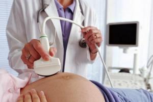
The difficulty is that it is not detected in the early stages of pregnancy. A doctor can only assume this disease if the mother is diagnosed. Therefore, the initial signs of fetal hypoxia are determined only with special equipment for examinations. In particular, ultrasound and Doppler (a technique for detecting a fetal heartbeat) are used for this purpose.
- Self-diagnosis
Many mothers, worried about the condition of their baby, want to know how to determine fetal hypoxia on their own and at what time it can be done. This becomes possible only at 18 weeks or even later, when the baby’s movements are felt. The woman should be observant during this period and notice how and when the baby usually moves. The first sign of hypoxia is a decrease in its activity, movements become rare, sluggish, and barely noticeable. If such a symptom is present, you must definitely tell the doctor who is observing you about it.
- Laboratory diagnostics
A decrease in the child’s activity in the womb may be a sign of other abnormalities in its development. To confirm or refute the diagnosis, additional examination is required, which can detect more obvious signs of hypoxia in the fetus.
- Ultrasound: physical data do not correspond to the norm (weight and size are smaller), there is a delay in development, premature maturation of the placenta, the presence of too thin or too thick walls;
- Doppler measurements: blood flow disturbances in the uterine arteries and placenta, bradycardia (low heart rate).
- Cardiotocography (indicated in documents as CTG and performed only after the 30th week) often gives incorrect results, so it is done several times to confirm the data.
- Sometimes amnioscopy is used, which reveals the state of amniotic fluid, which provides fairly reliable data on whether it is possible to talk about hypoxia in a particular case: in the presence of this disease, they become cloudy.
To be more confident in the diagnosis, a pregnant woman may be prescribed a number of blood tests - hormonal and biochemical. If they detect a high concentration of enzymes, fat oxidation products, such results will also indicate a disease.

Treatment of intrauterine hypoxia
Timely diagnosis and the absence of indications for immediate cesarean section (in which cases it is done, read) will allow a course of treatment for hypoxia during pregnancy to reduce the risk of illness in the baby after birth.
To achieve this, a whole range of activities is carried out:
- The cause of the development of oxygen starvation of the fetus is determined.
- If possible, it is eliminated immediately. If the issue is a woman’s failure to comply with basic recommendations during pregnancy, they explain to her how this could all end. Regular walks, good nutrition, good sleep, and the absence of bad habits can save your baby from this problem. If the cause is some kind of disease of the mother and its treatment is possible in this situation, all possible measures are taken to get rid of it.
- If necessary, bed rest is prescribed, which helps improve blood supply to the uterus.
- Medicines are prescribed: no-shpu, bricanil, suppositories with papaverine, ginipral (they reduce uterine contractility); reopolyglucin, aspirin, chimes (restore blood circulation); Essentiale-Forte, lipostabil (improves cell permeability to oxygen); , glutamic and ascorbic acids, glucose solution (to restore impaired metabolism);
- It is recommended to drink water enriched with oxygen.
Sometimes even complex therapy for fetal hypoxia is ineffective. And if the fetus has already reached viability, doctors decide to perform an emergency delivery. In order not to lead to such an outcome and not to risk the health of your own baby, it is better to warn him in advance with preventive measures.
Prevention

Effective prevention of hypoxia during pregnancy includes a whole range of measures that will help avoid the disease and its consequences. The expectant mother should lead a healthy lifestyle:
- get rid of bad habits;
- spend a lot of time in fresh, clean air (preferably away from chemical plants and highways);
- visit a doctor regularly;
- keep the doctor informed of all your past and present diseases;
- eat right, including iron-rich foods in your diet;
- master breathing exercises;
- have a good rest;
- sleep at least 8–9 hours a day;
- do not overwork;
- avoid stress and nervous experiences.
All these recommendations on how to avoid fetal hypoxia during pregnancy are very important to follow for absolutely all women, regardless of whether they are at risk or not. By following them, you can bear a strong baby without any developmental disabilities. If you take this lightly, dangerous consequences cannot be avoided.
Dangerous consequences of hypoxia during pregnancy
The consequences of different forms of fetal oxygen starvation may not be the same.
Chronic hypoxia
Late diagnosis and lack of treatment of the pathology leads to prolonged oxygen starvation, which is called chronic fetal hypoxia. The consequences present the saddest picture:
- the formation of fetal organs is disrupted;
- deviations in embryo development;
- damage to the central nervous system;
- growth retardation (impaired physical development);
- poor adaptation of the child to life outside the womb.
Newborns with chronic hypoxia will have poor appetite, will be restless, and the autonomic nervous system will be severely damaged.
Acute hypoxia
Acute fetal hypoxia does not predispose to therapeutic intervention. If the child is already viable, an emergency operation is performed to remove the fetus. If this is not done in time, prolonged oxygen starvation will lead to the death of brain cells and (respiratory impairment).
A pregnant woman must take care of both the baby and herself, since his condition depends on her state of health, diet and diet. If you want to carry, give birth and raise a healthy, strong baby, then save him from intrauterine oxygen starvation.
– intrauterine syndrome, characterized by a complex of changes in the fetus caused by insufficient oxygen supply to its tissues and organs. Fetal hypoxia is characterized by disorders of vital organs, primarily the central nervous system. Diagnosis of fetal hypoxia includes cardiotocography, Dopplerometry of the uteroplacental circulation, obstetric ultrasound, and amnioscopy. Treatment of fetal hypoxia is aimed at normalizing uteroplacental blood flow and improving blood rheology; sometimes this condition requires the woman to give birth early.

General information
It is registered in 10.5% of cases of the total number of pregnancies and births. Fetal hypoxia can develop at different stages of intrauterine development, characterized by varying degrees of oxygen deficiency and consequences for the child’s body. Fetal hypoxia, which develops in the early stages of gestation, causes defects and slower development of the embryo. In late pregnancy, hypoxia is accompanied by fetal growth retardation, damage to the central nervous system, and a decrease in the adaptive capabilities of the newborn.
Causes of fetal hypoxia
Fetal hypoxia can be a consequence of a wide range of unfavorable processes occurring in the body of the child, mother or placenta. The likelihood of developing hypoxia in the fetus increases with diseases of the maternal body - anemia, cardiovascular pathology (heart defects, hypertension), diseases of the kidneys, respiratory system (chronic bronchitis, bronchial asthma, etc.), diabetes mellitus, toxicosis of pregnancy, multiple pregnancy, STIs . Alcoholism, nicotine, drug addiction and other types of maternal addiction have a negative impact on the oxygen supply to the fetus.
The danger of fetal hypoxia increases with disorders of the fetal-placental circulation caused by the threat of miscarriage, post-term pregnancy, umbilical cord pathology, fetoplacental insufficiency, abnormalities of labor and other complications of pregnancy and the birth process. Risk factors for the development of intrapartum hypoxia include hemolytic disease of the fetus, congenital malformations, intrauterine infection (herpetic infection, toxoplasmosis, chlamydia, mycoplasmosis, etc.), repeated and tight entanglement of the umbilical cord around the baby’s neck, long-term compression of the head during childbirth.
In response to hypoxia in the fetus, the nervous system primarily suffers, since nervous tissue is most sensitive to oxygen deficiency. Starting from 6-11 weeks of embryo development, lack of oxygen causes a delay in brain maturation, disturbances in the structure and functioning of blood vessels, and a slowdown in the maturation of the blood-brain barrier. The tissues of the kidneys, heart, and intestines of the fetus also experience hypoxia.
Minor fetal hypoxia may not cause clinically significant damage to the central nervous system. With severe fetal hypoxia, ischemia and necrosis develop in various organs. After birth, a child who has developed under hypoxic conditions may experience a wide range of disorders - from neurological disorders to mental retardation and severe somatic abnormalities.
Classification of fetal hypoxia
Based on the time course and rate of occurrence, acute and chronically developing fetal hypoxia are distinguished.
The occurrence of acute fetal hypoxia is usually associated with anomalies and complications of labor - rapid or protracted labor, compression or prolapse of the umbilical cord, prolonged compression of the head in the birth canal. Sometimes acute fetal hypoxia can develop during pregnancy: for example, in the case of uterine rupture or premature placental abruption. In acute hypoxia, dysfunction of the vital organs of the fetus rapidly increases. Acute hypoxia is characterized by an increase in the fetal heart rate (more than 160 beats per minute) or a decrease in heart rate (less than 120 beats per minute), arrhythmia, deafness of tones; increased or decreased motor activity, etc. Fetal asphyxia often develops against the background of acute hypoxia.
Chronic hypoxia is caused by prolonged moderate oxygen deficiency, under which the fetus develops. With chronic oxygen deficiency, intrauterine hypotrophy occurs; in case of depletion of the compensatory capabilities of the fetus, the same disorders develop as in the acute version of the course. Fetal hypoxia can develop during pregnancy or childbirth; Hypoxia that occurs in a child after birth due to hyaline membrane disease, intrauterine pneumonia, etc. is considered separately.
Taking into account the compensatory and adaptive capabilities of the fetus, hypoxia can take on compensated, subcompensated and decompensated forms. Since under unfavorable conditions the fetus experiences not only hypoxia, but also a whole complex of complex metabolic disorders, in world practice this condition is defined as “distress syndrome”, which is divided into prenatal, developed during childbirth and respiratory.
Manifestations of fetal hypoxia
The severity of changes that develop in the fetus under the influence of hypoxia is determined by the intensity and duration of the oxygen deficiency experienced. The initial manifestations of hypoxia cause an increase in heart rate in the fetus, then a slowdown and muffled heart sounds. Meconium may appear in the amniotic fluid. With mild hypoxia, the motor activity of the fetus increases, with severe hypoxia, movements are reduced and slowed down.
With severe hypoxia, the fetus develops circulatory disorders: there is a short-term tachycardia and a rise in blood pressure, followed by bradycardia and a decrease in blood pressure. Rheological disturbances are manifested by thickening of the blood and release of plasma from the vascular bed, which is accompanied by intracellular and tissue edema. As a result of increased fragility and permeability of the vascular walls, hemorrhages occur. A decrease in vascular tone and slower blood circulation leads to ischemia of organs. With hypoxia, acidosis develops in the fetal body, the balance of electrolytes changes, and tissue respiration is disrupted. Changes in the vital organs of the fetus can cause intrauterine death, asphyxia, and intracranial birth injuries.
Diagnosis of fetal hypoxia
Suspicion that the fetus is experiencing hypoxia may arise when there is a change in its motor activity - restless behavior, increased and frequent movements. Prolonged or progressive hypoxia leads to weakening of fetal movements. If a woman notices such changes, she should immediately contact a gynecologist who is caring for the pregnancy. When listening to the fetal heartbeat with an obstetric stethoscope, the doctor evaluates the frequency, sonority and rhythm of heart sounds, and the presence of murmurs. To detect fetal hypoxia, modern gynecology uses cardiotocography, fetal phonocardiography, Doppler, ultrasound, amnioscopy and amniocentesis, and laboratory tests.
During cardiotocography, it is possible to track the fetal heart rate and its motor activity. By changing the heartbeat depending on the rest and activity of the fetus, its condition is judged. Cardiotocography, along with phonocardiography, is widely used in childbirth. Dopplerography of uteroplacental blood flow examines the speed and nature of blood flow in the vessels of the umbilical cord and placenta, the disruption of which leads to fetal hypoxia. Ultrasound-guided cordocentesis is performed to collect cord blood and study acid-base balance. An echoscopic sign of fetal hypoxia can be a detectable delay in its growth. In addition, during obstetric ultrasound, the composition, volume and color of amniotic fluid is assessed. Severe polyhydramnios or oligohydramnios may signal trouble.
Childbirth during chronic fetal hypoxia is carried out using cardiac monitoring, which allows timely application of additional measures. In case of acute hypoxia that develops during childbirth, the child requires resuscitation care. Timely correction of fetal hypoxia, rational management of pregnancy and childbirth help to avoid the development of gross disorders in the child. Subsequently, all children who developed under hypoxic conditions are observed by a neurologist; Often they need the help of a psychologist and speech therapist.
Complications of fetal hypoxia
Severe fetal hypoxia is accompanied by severe multiple organ dysfunctions in the newborn. With hypoxic damage to the central nervous system, perinatal encephalopathy, cerebral edema, areflexia, and convulsions can develop. From the respiratory system, posthypoxic pneumopathy and pulmonary hypertension are noted; cardiovascular disorders include heart and vascular defects, ischemic endocardial necrosis, etc.
The effect of fetal hypoxia on the kidneys can manifest itself as renal failure, oliguria; on the gastrointestinal tract - regurgitation, vomiting, enterocolitis. Often, due to severe perinatal hypoxia, a newborn develops DIC syndrome and secondary immunodeficiency. Asphyxia of newborns in 75-80% of cases develops against the background of previous fetal hypoxia.
Prevention of fetal hypoxia
Preventing the development of fetal hypoxia requires a woman to prepare responsibly for pregnancy: treatment of extragenital pathology and diseases of the reproductive system, giving up unhealthy habits, and a balanced diet. Pregnancy management should be carried out taking into account risk factors and timely monitoring of the condition of the fetus and woman. Preventing the development of acute fetal hypoxia lies in the correct choice of method of delivery and prevention of birth injuries.
We all know that during pregnancy, a woman's thoughts rush in one direction. She dreams of a future baby, of a happy life together, she already cares about his condition and comfort and wants the baby to be born strong, healthy and on time.
In order for the fetus to develop fully during pregnancy and the birth to take place safely, all processes in both organisms - the child's and the mother's - must proceed normally, as expected. Any violation can affect the baby's condition. And such a situation can arise for many reasons. Often pregnant women are diagnosed with fetal hypoxia. And this is a serious reason to think and act.
What's happening?
The word "hypoxia" means lack of oxygen. That is, when we talk about fetal hypoxia, this means that the baby does not receive enough oxygen from the mother’s body, and oxygen starvation of the fetus occurs, as doctors say. This condition can develop during pregnancy (and then a diagnosis of chronic hypoxia is made) or directly during childbirth (we are talking about acute hypoxia).
What happens when there is not enough oxygen? Of course, the baby begins to choke. But not right away. First, a number of disorders occur in his small body, the consequences of which, if hypoxia is not detected and therapeutic measures are not taken on time, can be irreversible.
Lack of oxygen in the early stages of pregnancy (when the formation and formation of organs and systems occurs) can provoke disruption of the development of the embryo, including anomalies and injuries. And in the later stages, the child’s central nervous system and physical development suffer: growth is delayed, the newborn adapts less well to the new environment, and may have physical and mental abnormalities. Children born with hypoxia have disorders of the autonomic nervous system, muscle hypertonicity, the baby is restless, capricious, eats and sleeps poorly. Such a child should be under constant supervision of a neurologist.
When the fetus experiences a lack of oxygen, all its organs and systems begin to work in increased mode, trying to obtain the vital gas. This is possible due to the increased compensatory capabilities of the small organism. The woman feels this activation through the increased mobility of the baby. But this may not last long. And if the normal supply of oxygen is not restored and the metabolism is not normalized in time, depression soon sets in - the child becomes quiet, because without oxygen he can no longer move. The consequences of this condition may be irreversible. Therefore, if after suddenly increased activity your baby suddenly freezes (you feel no more than 3 movements per hour), you should consult a doctor immediately! Hypoxia can be detected most reliably through additional studies: cardiotocography and Doppler.
Why does this happen?
Oxygen is supplied to all our organs and systems along with blood. Transports oxygen, and without iron it is not produced. That is, with (iron deficiency), the production of hemoglobin and, accordingly, the flow of oxygen into the blood and further throughout the body naturally decreases. However, a lack of iron in the mother's blood is not the only cause of hypoxia.
During pregnancy, the volume of circulating blood in the mother’s body increases significantly, because through the placenta it nourishes the fetus. If the uteroplacental exchange deteriorates, the embryo cannot receive the required amount of all nutrients, including oxygen, supplied to it with maternal blood. Metabolic disorders between mother and fetus occur with placental insufficiency. It also blocks the flow of oxygen to the fetus during pregnancy, since nicotine constricts blood vessels and blood circulation is impaired. It is also known that tobacco smoke penetrates to the embryo through the placenta, and it ends up in a smoke screen - how can you not suffocate... It does not have the best effect on blood vessels and...
In general, the development of hypoxia can be provoked by a number of diseases (especially chronic diseases of women) and disorders in the organisms of the fetus and mother and in the placenta:
- cardiovascular diseases of a pregnant woman;
- anemia;
- pulmonary diseases (respiratory tract);
- deep;
- gestosis;
- post-maturity;
- polyhydramnios;
- multiple births;
- violations during;
- threat ;
- pathology of the placenta and umbilical cord;
- anomalies of labor;
- intrauterine infections, intoxication;
- hemolytic disease of the fetus;
- prolonged compression of the head during childbirth and others.
Thus, hypoxia should be considered as a condition caused by a complex of changes in the body of the mother and child.
How to treat?
If a pregnant woman is diagnosed with hypoxia, she may be hospitalized in a hospital to ensure complete rest and provide the necessary treatment. Although it is quite possible that treatment can be done at home with a visit to a clinic or hospital. The doctor must find out what disease caused the development of hypoxia and prescribe appropriate treatment.
Therapy is carried out comprehensively. However, if no positive dynamics are observed and the condition of the fetus worsens, the issue of performing a cesarean section is considered (but this is only for a period of at least 3 months).
How to prevent it?
Approximately 10.5 percent of women are diagnosed with fetal hypoxia. However, in order not to be on their list, you just need to adhere to a certain lifestyle during pregnancy.
The most important thing is not to smoke or drink alcohol. If possible, breathe only clean air. That is, if you live in a very gas-polluted area, move to a cleaner area for this time. Ventilate the room in which you live as often as possible. Spend every day outdoors, but don’t forget about proper rest.
Nutrition and prevention of iron deficiency anemia are of paramount importance.
Of course, even a healthy lifestyle and good nutrition cannot be a 100% guarantee that hypoxia will not develop during pregnancy. But it will significantly increase your chances of preventing it. In addition, regular examinations by a gynecologist and consultations with a doctor will help to identify something wrong in time.
Acute fetal hypoxia
A few more words about the lack of oxygen experienced by the child directly during childbirth - acute fetal hypoxia. This condition can arise for a number of reasons: a very rapid or very prolonged labor, when the baby stuck in the birth canal simply cannot breathe; entanglement of the fetus with the umbilical cord; premature placental abruption. All this leads to fetal asphyxia (suffocation).
If acute hypoxia develops, the doctor delivering the baby monitors the condition of the fetus, in particular, conducts cardiac monitoring, tracking cardiac activity. A prerequisite for this may be cloudy greenish waters: this means that meconium has entered them. This criterion can be taken into account only in case of cephalic presentation of the fetus. In addition, acute hypoxia can be judged by tests of amniotic fluid and fetal blood tests (based on pH level).
A prolonged increasing state of hypoxia during childbirth is an indication for an emergency cesarean section.
But it should be understood that even acute hypoxia has its roots in the period of gestation. And if the violations and changes that arise at this time are identified in advance, then many troubles can be avoided.
Especially for- Elena Kichak
Intrauterine fetal hypoxia- a pathology characterized by a lack of oxygen in the body of the unborn child. Deficiency of this chemical element leads to disturbances in cell metabolism, and subsequently to their death. Fetal hypoxia is the cause of intrauterine growth retardation and development of the unborn child, congenital anomalies of the central nervous system and the death of the baby.
Diagnosis of oxygen starvation of the fetus is one of the main tasks of every appointment with an obstetrician-gynecologist. Timely identified pathology ensures the selection of the correct treatment and the prevention of severe complications.
Fetal blood supply
In the womb of the mother's body, the lungs of the unborn child are in a collapsed state and do not participate in the act of breathing. The supply of oxygen and the removal of carbon dioxide is ensured by the vessels of the umbilical cord. It contains two arteries carrying venous blood (poor in oxygen) and one vein carrying arterial blood (rich in oxygen).One end of the umbilical cord faces the fetus and is fixed in its anterior abdominal wall. The opposite part of the arteries and veins connects with the vessels of the placenta. Then they decrease in diameter and flow into the capillaries of the villi. This place is the point of intersection of the uterine (maternal) and placental (fetal) vessels. It is here that gas exchange takes place between the blood of the expectant mother and the child.
A pathological process in any of the listed areas leads to hypoxia of the unborn child. Most often, damage occurs at the level of the uterine and placental vessels at their junction. Also, fetal hypoxia may be associated with extragenital pathology of the mother, leading to insufficient blood supply.
Classification
Doctors classify fetal hypoxia according to several criteria. According to the time of development of the process, acute and chronic types of pathology are distinguished.Acute fetal hypoxia develops over a rapid period of time, from several minutes to several hours. It is usually associated with placental abruption or vascular thrombosis. In the absence of medical care, acute oxygen starvation often ends in intrauterine fetal death.
Chronic fetal hypoxia is a long-term process that develops over several days or weeks. This type of oxygen starvation usually occurs against the background of concomitant pathologies - anemia, gestosis, diabetes mellitus. Constant hypoxia is the cause of intrauterine growth retardation (hypotrophy) and pathologies of the central nervous system of the unborn child.
Depending on the level of damage, the following types of hypoxia are distinguished:
Hypoxic. Occurs with pathologies of the blood vessels of the placenta or uterus. Also, this type of oxygen starvation may be associated with concomitant maternal diseases.
Hemic. Occurs with pathologies of the fetal blood system, when its red blood cells cannot bind oxygen. The most striking example of this type of hypoxia is hemolytic disease.
Circulatory. The type of oxygen starvation is associated with damage to the vessels of the umbilical cord or fetus. With this type of pathology, the placental blood supply is not impaired. Circulatory hypoxia occurs with congenital heart defects of the fetus, as well as with compression of the arteries and veins of the umbilical cord.
Fabric. A rare type of fetal hypoxia associated with metabolic disorders in the body of the unborn child. Typically, tissue oxygen starvation occurs with congenital pathologies of enzyme systems.
Depending on the severity of the course, there is a third classification of fetal hypoxia. The functional or compensated form of oxygen starvation is the easiest; it does not lead to disturbances in the body of the unborn child.
The metabolic or subcompensated form is characterized by metabolic disorders and the accumulation of harmful products. However, this type of hypoxia is reversible. With timely provision of medical care, the birth of a completely healthy child is possible.
The destructive or decompensated form is the most severe fetal hypoxia. It is accompanied by irreversible processes in the body of the unborn child, pathologies in the central nervous system and other anatomical structures.
Doctors also distinguish primary hypoxia, which occurs before the 16th week of pregnancy, and secondary hypoxia, which develops at a later date.
Causes
There are many reasons that cause intrauterine fetal hypoxia. The most common of them include the following factors:Gestational hypertension (late gestosis)
This pathology occurs due to improper development of the uteroplacental vessels after 20-22 weeks of pregnancy. To restore blood flow, a woman’s body reflexively increases blood pressure. For some time, this measure is effective.However, with an increase in blood pressure, spasm of the blood vessels of the uterus and placenta is observed. A decrease in the diameter of the arteries leads to a decrease in blood flow in them and to chronic fetal hypoxia.
Symptoms of late gestosis in pregnant women include increased blood pressure, swelling and the appearance of protein in the urine. Typically, the first signs of arterial hypertension appear after 32 weeks of gestation. An earlier onset of clinical manifestations indicates a severe course of the pathology.
Premature abruption of a normally located placenta
Premature placental abruption most often occurs during childbirth, but it can occur throughout pregnancy. This pathology is the most common cause of acute fetal hypoxia.The pathogenesis of placental abruption is associated with its improper attachment, structural abnormalities, and increased emotional or physical stress. Sometimes this disorder occurs due to a lack of progesterone. Detachment of more than half the area of the placenta leads to immediate fetal death.
Symptoms of premature placental abruption are uterine bleeding and cramping pain in the lower abdomen. If these signs are present, the expectant mother should immediately seek medical help.
Anemia
Anemia is a lack of hemoglobin in a unit of blood. Most often, expectant mothers are prone to developing the iron deficiency type of this pathology. Less commonly, anemia occurs due to a lack of vitamin B12, folic acid, bleeding or a disease accompanied by the breakdown of red blood cells (malaria).The main consequence of anemia is chronic fetal hypoxia. The main symptoms of maternal pathology include dizziness, nausea, weakness, pale skin, and fainting.
Infectious diseases
Viral and bacterial diseases are a risk factor for intrauterine fetal hypoxia. Some infections affect the homeostasis system, causing pathologies of the blood coagulation system. Diseases contribute to the formation of microthrombi that clog the lumen of the uterine and placental vessels.Also, the infectious disease itself can cause a state of intoxication, which contributes to a decrease in oxygen in the blood. Prolonged high fever causes fetal hypoxia.
Multiple pregnancy
When carrying twins or triplets, the likelihood of intrauterine fetal hypoxia increases significantly. This feature is associated with an increase in oxygen demand due to distribution between several fruits.Fetal movements / when should you worry?
Degrees
During an ultrasound examination using a Doppler probe, doctors distinguish three degrees of fetoplacental insufficiency:- Type 1a of fetal oxygen starvation is accompanied by impaired blood supply in the uteroplacental vessels;
- Type 1b of oxygen starvation of the unborn child is characterized by pathologies of blood flow in the fetal-placental area;
- Stage 2 fetal hypoxia is characterized by impaired blood flow in both systems, but they are in a state of compensation;
- Stage 3 oxygen starvation of the fetus is accompanied by a violation in any of these systems, accompanied by a threat to the life of the fetus.
Symptoms
Symptoms of fetal hypoxia are subjective; they cannot speak with absolute certainty about the presence of pathology. That is why expectant mothers should not skip routine examinations and consultations with an obstetrician-gynecologist.Mild and moderate fetal hypoxia usually does not manifest itself in any way. In the later stages of pregnancy, the expectant mother may notice a change in the nature of fetal movements. In the acute form of the pathology, the baby begins to move intensively; in the chronic type of hypoxia, his activity may be reduced.
A severe decompensated form of oxygen starvation of the fetus is often manifested by intrauterine growth retardation and development of the unborn child. That is why the expectant mother may notice a slow increase in abdominal girth and a lag in the height of the uterine fundus from the period of pregnancy.
To independently diagnose fetal hypoxia, the expectant mother can try to listen to its heartbeat using a phonendoscope. This method is possible only after the 20th week of pregnancy. The normal heart rate of an unborn child ranges from 120 to 160 beats per minute.
An increase in heart rate often accompanies acute fetal hypoxia. A pulse of less than 120 beats per minute can be observed with chronic oxygen starvation of the unborn child.

Diagnostics
To diagnose the condition of the unborn child, various instrumental research methods are used. The simplest of them is ultrasound. Using ultrasound, the doctor can indirectly judge the presence or absence of fetal hypoxia.Ultrasound equipment allows you to see the structure of the placenta, detect areas of detachment, its aging, and measure the thickness of the organ. Specialists can also visualize the fetus’s body for the presence of congenital pathologies of the heart and blood vessels, as well as the correspondence of its size to the gestational age.
Gives a more accurate picture of the state of the blood supply to the fetus. This diagnostic method is based on the presence of a special sensor that reads information about the direction and speed of fluid flow in the vessels.
Using Doppler, doctors can visualize blood flow in all vessels of the uterus, placenta, umbilical cord and fetus. The instrumental research method makes it possible to determine the degree of fetal hypoxia and make a prognosis about the further course of pregnancy.
Cardiotocography is a method of instrumental diagnosis of the condition of the fetus. CTG allows us to indirectly judge the presence or absence of hypoxia in the unborn child. The principle of operation of this equipment is to record the baby’s heart rate in response to stimuli.
In the presence of hypoxia, the heart rate rhythm is monotonous, the average pulse is less than 120 or more than 160 beats per minute. Normally, the child should not experience decelerations - periods of heart rate decrease by 30 or more beats per minute for a period of more than 30 seconds.

Treatment
Treatment of fetal hypoxia depends on the cause that caused it. If there is a subcompensated and decompensated form of oxygen starvation of the unborn child, the woman requires hospitalization. According to strict indications, premature delivery is possible.The basic principles of treatment of intrauterine fetal hypoxia is the resumption of normal blood circulation. For this purpose, the expectant mother is given medications that expand the lumen of blood vessels (Eufillin). Pregnant women are also advised to take drugs that improve metabolism in tissues ().
In the presence of uterine hypertonicity, the use of myotropic drugs is indicated (,). Also, all expectant mothers are recommended to take B vitamins, which improve the rheological properties of the blood.
If the expectant mother has a specific disease, she is indicated for special therapy aimed at treating or compensating for it. For deficiency anemia, iron supplements, folic acid and vitamin B12 should be taken. For the treatment of gestational arterial hypertension, the use of Methyldopa and.
Consequences
Acute fetal hypoxia is a risk factor for intrauterine fetal death. A chronic type of oxygen deficiency can cause various consequences. Most often, severe fetal hypoxia is the cause of delayed growth and development. The likelihood of congenital pathologies of the central nervous system also increases. Cells of the brain and spinal cord are most sensitive to oxygen deficiency.Children suffering from hypoxia during intrauterine life may differ from their peers. This pathology causes mental and mental retardation and brain diseases. Quite often, after birth, such children have a restless character and are difficult to train in the future.

Prevention
In order to prevent fetal hypoxia, the expectant mother is recommended to lead a healthy lifestyle. A pregnant woman should avoid smoking and alcohol, emotional stress and heavy physical labor. Her diet should include a variety of healthy foods, enriched with all vitamins and minerals.The basis for preventing oxygen starvation of the fetus is pregnancy planning. Before conceiving, the expectant mother is recommended to compensate for all chronic pathologies and be tested for sexually transmitted diseases.
Hypoxia during childbirth
Fetal asphyxia- an acute state of lack of oxygen during childbirth with preservation of cardiac activity; this term is synonymous with hypoxia. Usually the pathology occurs due to disruption of uterine contractions, trauma, or a clinically narrow pelvis. Fetal hypoxia during childbirth can also be caused by improper use of medications.Fetal hypoxia at birth is diagnosed using a CTG machine. Its consequences include possible complications on the central nervous system and potential fetal death. To treat oxygen starvation of the unborn child, contractions should be normalized or an emergency cesarean section should be performed.