It happens that at 32-33 weeks of pregnancy, the expectant mother learns about the wrong presentation of the fetus. At an earlier stage, the baby is still quite mobile, and it is premature to worry about his position. But at the 34th week, it is already possible and necessary to take measures. There is a special gymnastics that helps to solve the problem of oblique, transverse or pelvic presentation of the fetus. The exercises are mostly simple and suitable for independent home practice.
If a woman has experience in classes, then you can add to the simplest gymnastics and other poses with the initial position on all fours, with support on knees and elbows - various variations of the Cat-Cow ligament, especially in dynamics, Dolphin pose, Downward Dog pose and her modifications, movements of the pelvis "figure eight" in a standing position, etc.
All these asanas and exercises that are well known to us from the practice of yoga for pregnant women will help the child to turn his head down, and it is this position that is the safest and most natural for both mother and baby. Let's take a look at some of the exercises that have proven effective in oblique, transverse and breech presentation. Perform all exercises, alternate or select the most appropriate for your health and fitness level. Choose a time when your child is active. Exercise on an empty stomach!
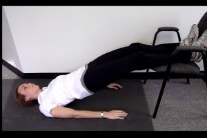
Transverse and breech presentation of the fetus: exercises with the position of the pelvis above the chest
Do these exercises for 10-15 minutes two to three times a day. The idea behind this cross or breech presentation is to raise and fix your hips above your shoulders, giving your baby extra space and time to make the desired maneuver. As a result, gravity itself will help the baby to turn his head towards the exit from the uterus.
 |
This is one of the main exercises for transverse presentation of the fetus. You will need a chair and pillows or blankets. Lie on the floor with your feet on a chair. |
 |
Lift your pelvis up and place pillows or blankets under your pelvis (by yourself or with someone's help) so that the body and hips are at an angle of 45 degrees to the floor, forming a straight line. |
 |
The starting position is on all fours. |
 |
Place pillows or blankets under your knees and head. Lower your head to the floor and relax. |
 |
You can do this exercise without pillows, just on a rug. |
Transverse and breech presentation of the fetus: exercise 4, Dolphin pose
Dolphin Pose requires a certain amount of physical fitness. It is suitable for women who practiced yoga before pregnancy and then continued to keep fit with yoga for pregnant women.
 |
As you inhale, push the body forward so that your head protrudes beyond the hands that are locked in the wrist lock. |
 |
As you exhale, bring your body back, essentially into Downward Dog Pose, resting on your forearms. The abdomen is relaxed. Do until tired, then rest in exercise 2. |
This pose is also for those pregnant women who have done yoga before, and now continue by attending yoga classes for pregnant women. Don't risk trying it now: your neck may not be ready for the pressure that is placed on it in the Shoulderstand, even in its adapted version.
With a transverse presentation of the fetus, it is also very useful (preferably in the presence and with the help of someone close to you) to climb onto the sofa with your knees, and lower your forearms to the floor and lean on them. Your hips and knees should be about 10-20 centimeters above your shoulders. It turns out something like strong modified posture of the Downward-facing Dog with support on the forearms.
Relax your stomach, neck, and head. Hold the position for 30-60 seconds. Then slowly slide off the couch - first lower one knee down, then the other. Get into a sitting position and catch your breath. Repeat. This exercise should be done twice a day for several days.
You can also place a warm heating pad on the lower abdomen to show the baby a more comfortable place for the head. I have also heard more than once that a child can turn around, moving his head towards the light: for this, they shine a flashlight in the lower abdomen, in the pubic area. Also ask your husband or mother to bend over to the pubis and call the baby from there. Sometimes they even use a homemade voice amplifier for this - a tube rolled out of cardboard.
Transverse and breech presentation of the fetus: exercises in a position on all fours
The purpose of the exercises in this group is to relax the belly and the baby, encouraging him to take the correct position with his smooth but prolonged movements. Perform this type of exercise for 5-10 minutes, relaxing the abdomen, pelvic floor and perineum, as if directing inhalation and exhalation there.
Transverse and breech presentation of the fetus: exercises in a standing position, sitting on a fitball and lying
Transverse and breech presentation of the fetus: exercise 10, lunge with a leg on a chair in dynamics
An asymmetrical pelvic position can be very helpful in encouraging your toddler to get into an optimal position.
 |
Stand next to a chair, put one leg on it, turning its foot perpendicular to the back. The lower foot is parallel to the back of the chair |
 |
Move the pelvis towards the back of the chair, then return to the starting position, continue to move gently in the modified lunge, then do the exercise on the other leg. |
You can do a similar exercise on a ladder. Or get on all fours, straighten your torso and lift one knee. Resting on the foot of your front foot, perform a lunge in the same way as in the described chair exercise. Change your leg.
In addition, every day, from morning to evening, you need to monitor your posture. Pay attention to how you sit, stand, and walk. By slouching, you are not helping your baby to turn around. Open your shoulders, push your chest forward and up, try to smooth out the lumbar lordosis by tucking the tailbone towards you, and pushing the lower ribs and sides of the waist backward. Keep your pelvic floor toned.
In general, the posture should be the same as during ... yoga classes :-). This gives the uterus the ability to take a shape that encourages the baby to turn in the right direction.
And move more! A few short walks each day should become a habit. Shake your pelvis more often, like an oriental dancer. Draw eights with your pelvis - standing and resting on your knees and elbows, relaxing your stomach. The more you move your hips, the easier it is for your baby to get into the optimal position in your abdomen and prepare for childbirth.
And after the baby turns around, it is worth fixing the stomach with a special bandage so that he does not change his mind again.
The cost of an individual yoga class for pregnant women in order to change the presentation of the fetus: RUB 3200
and receive new issues of the blog directly to your inbox!
Content
The situation when a child is in breech presentation in a pregnant woman in the last stages is considered rare - there are about 5 women with this deviation per 100 pregnant women. This explains the fact that many pregnant women do not know what the breech presentation of the fetus, the incorrect position of the baby's head in the uterus, can threaten the child and the pregnant woman herself during childbirth, what pathologies occur in the baby if the birth is not carried out skillfully and competently. In other cases, the pelvic position of the fetus is an indication for caesarean section, as the safest method of childbirth.
What is breech presentation of the fetus
During the entire period of pregnancy, the embryo changes its position in the uterus several times. Gynecologists consider these movements to be a normal process until the last period of pregnancy, when, in most cases, the fetus takes a position with the head down, which is considered the correct presentation for natural childbirth. The fetal head is the most voluminous part of the body, therefore, during normal childbirth, when the head passes through the perineum, the rest of the body follows it inertially, without causing problems during delivery.
The situation when, after 30 weeks of pregnancy, an anatomical breech or foot presentation of the fetus is recorded by an obstetrician can greatly complicate childbirth. The child first has legs or buttocks, which do not take up much volume, and only then a head is born, during the passage of which through the birth canal difficulties may arise, fraught with the threat of serious pathologies in the born baby.
Causes
If the fetus is in breech presentation in the last stages of pregnancy, then there are many reasons for this condition. Factors affecting abnormal presentation of the fetus are divided into three main groups:
- Dependent on the mother, or maternal. These include: a narrow pelvis that prevents the child from taking the correct position with the head to the pelvic floor, a history of fibroids or fibroids, ovarian tumors, hypoplasia, pathological abnormalities in the structure of the uterus.
- Caused by abnormalities in the development of the fetus, or fruit. These include: polyhydramnios, entanglement of the umbilical cord around the embryo, its too short length, hypoxia, hydro-, anen- and microcephaly of the fetus, twins or triplets according to the results of ultrasound.
- Placental, when the breech presentation of the child is facilitated by a low placenta previa and a high tone of the lower parts of the uterus, caused by various operations, scars, and frequent curettage of the uterine cavity. The fetus tries to take the upper position when its head is not pressed by the spasmodic muscles of the uterus.
Classification
There are several types of abnormal presentation of the fetus in the mother's pelvic ring:
- Fully breech presentation of the fetus, when the buttocks of the embryo are lowered down, and it bends the legs and presses the arms against the tummy.
- Leg presentation, when the embryo has one or two feet in the pelvic ring. Sometimes the knees of the fetus are there.
- Mixed presentation. In this case, the buttocks and one foot are on the pelvic ring, the second leg is straightened.
What is dangerous
A condition with a breech presentation recorded by obstetricians is dangerous for the risk of early termination of pregnancy, which interferes with the normal formation of the central nervous system and endocrine systems of the embryo. In the last stages of gestation, the formation of the medulla oblongata of the embryo occurs, and the pelvic position of the fetus can lead to a disruption of this process, causing cerebral edema in the newborn baby. Malformations can also be recorded, including heart failure, anomalies in the development of bones, muscles, central nervous system, genitals.
Does the abdomen drop with breech presentation
One of the most important signs that the fetus is in breech presentation is that the belly of a pregnant woman in the last stages does not lower, but is in an elevated state. Down the stomach "pulls" the head, which, after 30-32 weeks, descends to the pelvic ring. If the head is located on the upper segments of the uterus, and the buttocks, feet or knees of the fetus are below, then the stomach will not go down.
Diagnostics
A stable breech presentation is recorded by gynecologists starting from 32 weeks of gestation during a planned gynecological examination of the pregnant woman. At the bottom of the uterus, a large head is felt, the heartbeat is felt opposite the navel, and at the entrance to the womb, you can feel the sacrum, spine, soft, irregularly shaped parts of the child's body, in which the buttocks, heels, feet with toes are guessed. Based on the data of visual examination, the gynecologist or obstetrician fixes the abnormal position of the embryo.
The pregnant woman is prescribed the following additional procedures to confirm the diagnosis of abnormal presentation: examination of the child using a three-dimensional ultrasound scan, which gives a three-dimensional picture of the position of the embryo in the uterus, Doppler ultrasound and cardiotocography, allowing to assess the health of the internal organs of the embryo that has taken an incorrect presentation.

Pregnancy management with breech presentation of the fetus
The difference between the observation of a woman with a fixed breech or foot presentation of the fetus from the standard management of pregnancy becomes attempts to correct the pelvic position of the embryo. For this, the following methods are used:
- The woman is prescribed special gymnastics, in which she must make turns from one side to the other and lift the pelvis above the level of the head from a prone position. Charging has contraindications: exercises cannot be done with scars and scars on the uterus, low placenta previa, preeclampsia.
- If gymnastics does not help, then doctors can hospitalize the patient and attempt an external turn in stationary conditions. With an incorrect external turn, rupture of the placenta, membranes, rupture of amniotic fluid and premature birth can be caused.
Childbirth
To determine how the delivery will go with a breech presentation of the fetus, the pregnant woman is admitted to the hospital at 33 weeks of gestation. The decision on the method of childbirth is made based on an assessment of the patient's general condition, the way the baby is positioned in the bosom of the uterus, a history of diseases that can negatively affect the intrauterine development of the baby, age, blood pressure of the pregnant woman, the number of previous pregnancies of the expectant mother, her readiness to follow orders obstetrician.
Biomechanism of labor in breech presentation
The pelvic position of the embryo determines other obstetric methods of carrying out natural childbirth than the head. Since the buttocks are considered the largest part of the baby's body after the head, the baby will be born according to the following algorithm:
- The buttock is born first, which is closer than the other to the birth canal. It descends into the small pelvis, where an overturn and displacement of the buttocks are carried out on the finger, as a result of which it is pulled out with the end forward, leaving the birth canal.
- Then the baby's pelvic region is fixed at the end of the pubic arch, the baby's spine is strongly bent, and a second buttock is born.
- If the baby's legs are bent at the knees, then they are born at the same time as the buttocks. With the legs along the torso, the obstetrician waits for the next contractions of the woman in labor to pull the legs out of the birth canal.
- The baby's torso passes through the birth canal easily if, before this stage, the birth of the baby's buttocks and legs passed without complications.
- The baby's shoulders are born in turn, with a fixed point of fixation. At the same time, the handles are released.
- Then the head is born, passing the sharp end forward in the transverse dimension. From the moment the baby was born to the shoulders, to the removal of the head, no more than 10 minutes should pass, because the head squeezes the umbilical cord, the baby begins to suffocate from lack of oxygen.

- rupture of the placenta, early discharge of amniotic fluid, prolapse of the umbilical cord, fraught with the fact that the child may suffocate;
- throwing back the handles;
- traumatism of the spine and head of the baby, leading to cerebral hemorrhages;
- water entering the baby's lungs while the head is still in the birth canal.
Consequences for the child
With an incorrectly conducted natural childbirth in the condition of the pelvic placement of the baby, the consequences for him will be the most serious, up to the presence of serious pathologies at birth and death. Therefore, doctors recommend a cesarean section as the safest method of obstetrics, in which the child has a high probability of being born healthy and without developmental disabilities.
Video
Found a mistake in the text? Select it, press Ctrl + Enter and we'll fix it!Pregnancy calendar: 33 weeks
33rd week of pregnancy: fetal development, weight, movement
The rapid growth of the fetus at the end of pregnancy leads to the fact that he has to take a forced position in order to fit in a given volume of the intrauterine space. The baby's head is tilted towards the chest, arms and legs are crossed and brought to the body.
Restriction of fetal mobility contributes to its final location in the uterine cavity. The most favorable is the cephalic presentation of the fetus, when the head is located at the bottom, at the entrance to the pelvis. But its reverse position, with the pelvic end down, is also not a disaster and natural childbirth with proper obstetric assistance is quite possible. An exceptional case is only the transverse position of the fetus, which is an indication for a caesarean section.
The maturity of the fetus is such that if he has to be born prematurely, he will be quite able to survive with sufficient care, and does not even need special measures to improve breathing and nutrition. The only thing that babies born at the age of 33 weeks cannot cope with is the independent maintenance of a constant body temperature, because the development of subcutaneous tissue is still insufficient.
In premature babies (33 weeks of gestation), the cartilage of the auricles becomes denser, striation of the feet appears, the nail plates practically cover the nail beds. The thickness of the muscular layer of the vessels increases, the heart begins to work harder to ensure adequate blood flow.
In the lungs, the formation of alveoli is completed, however, due to some rearrangements, the formation of surfactant begins to decrease. This process stretches over the next 2 weeks, during which the risk of developing a distress syndrome, which often premature babies often suffer from, increases - 33 weeks, a rather dangerous period for the birth of a child.
By the end of the 33rd week, the length of the fetus is 44 cm, the weight is 2000 g, the diameter of the head is 82.0 mm, the diameter of the chest is 85.0 mm, the diameter of the tummy is 87.4 mm. The thickness of the placenta should be about 33.04 mm.
Are you concerned about a question about your pregnancy? Ask your obstetrician-gynecologist.
33rd week of pregnancy: a woman's feelings
 At the thirty-third week of pregnancy, a woman increasingly feels the approaching birth. Thoughts about an upcoming event disturb her and sometimes prevent her from falling asleep, sometimes her stomach pulls, frightening with the premature resolution of pregnancy. In addition to this, heartburn is often troublesome in the evenings. resulting from a physiological increase in the acidity of gastric juice, provoked by late and abundant meals, the use of sour, fatty foods. With a lack of calcium and the accumulation of excess fluid, the muscles of the hands periodically cramp. In the supine position, there is often a feeling of heaviness in the lower back, and sometimes even pain spreading along the thigh to the knee is a consequence of the compression of the growing uterus of the femoral nerve passing through the neighborhood.
Strong tension of the skin of the anterior abdominal wall is accompanied by the occurrence of itching, which is relieved by applying a moisturizer or cream for stretch marks. If you want to preserve the aesthetic appearance of the abdomen, wear a bandage even when you are at home and get up from the couch for only 5 minutes, just brew tea. In this case, the uterus is supported in an upright position, does not descend or stretch the lower abdomen.
The pressure of the uterus towards the diaphragm limits the mobility of the lungs, which causes mild shortness of breath. At the 33rd week of pregnancy, the lack of air forces a woman to spend a significant part of the time reclining and periodically take deep breaths. You will have to endure until the 36-37 weeks of pregnancy, when the baby's head sinks into the pelvis and the bottom of the uterus moves slightly downward.
At the 33rd week of pregnancy, even women who do not suffer from late toxicosis may notice some swelling of the legs in the ankle area, especially after walking or doing housework. Swelling of the lower extremities is associated with difficulty in the outflow of blood through the veins compressed by the large uterus. For the prevention of edema, it is recommended to rest, giving the legs a slightly elevated position. Women suffering from varicose veins. you should especially carefully follow the doctor's recommendations and wear compression hosiery at all times. By the way, the same bandage that supports the uterus in an elevated position helps to facilitate blood flow through the veins of the legs.
The ideal way to prepare your body for childbirth is to do yoga. It's okay if you haven't tried it yet. With the guidance of a competent and experienced instructor, you can get started right away. If, for some reason, you are unable to attend yoga classes at a specialized center, look in a store or on the Internet for video yoga lessons for pregnant women. With most of the exercises for beginners, you can do it yourself without harm to yourself and your child.
33rd week of pregnancy: discharge from the genital tract
 At the thirty-third week of pregnancy, a woman, as before, must monitor the nature of the discharge from the genital tract and, if leucorrhoea, mucus, pus or blood appears, take immediate action.
Mucous, mucopurulent, curdled leucorrhoea is a symptom of an infection of the genital tract and a reason for an extraordinary visit to a doctor and additional examination. The female reproductive tract should be healthy and free of infection. Disease-causing microbes can cause a baby to become infected during pregnancy or at birth.
Bloody discharge from the genital tract is a sign of placenta previa or detachment of a normally located placenta. Even if it is just a slight admixture of blood, regardless of whether the stomach hurts or not, you should immediately call an ambulance and go to the maternity hospital. Sudden tension in the uterus can cause severe bleeding and kill the baby.
The appearance of liquid, resembling diluted milk or slightly yellowish vaginal discharge, is alarming for the outpouring of amniotic fluid. Water can be poured out at once, in a stream and pose a threat to a woman. But in some cases, they leak and pour out in small portions, causing only some discomfort. Violation of the integrity of the membranes in the absence of labor during the day can lead to the development of intrauterine infection of the fetus.
33rd week of pregnancy: nutrition of the pregnant woman
Heartburn of pregnant women is a very unpleasant symptom, but sometimes the woman herself provokes its occurrence. Violation of the diet, late dinner, overeating, the use of spicy, sour, fatty, carbohydrate foods leads to an increase in the acidity of gastric juice and prolonged retention of food in the stomach, and the physiological weakness of the sphincter that isolates the stomach from the esophagus, and the pressure of the uterus on the abdominal organs become immediate the cause of the throwing of contents from the stomach into the esophagus.
What can help prevent heartburn?
- Frequent and fractional meals (5-6 times a day) and the last meal no later than 3-4 hours before bedtime.
- Refusal from fatty, fried, spicy, smoked products, coffee, strong tea, chocolate.
- Light dinner, mainly consisting of easily digestible proteins and carbohydrates, such as porridge and steamed cutlet.
- Moderate activity after eating. You should not immediately lie down on the sofa for a meal, it is better to take a walk or at least wash the dishes.
- Daily bowel movement.
- Wearing loose, non-abdominal clothing.
- Sleep on a bed with the head end raised.
If heartburn still persists, you should take measures to reduce the acidity of gastric juice. Baking soda, previously used by our mothers and grandmothers, is a barbaric method. The consumption of soda is accompanied by increased gas formation and only for a while reduces the acidity in the stomach. After about half an hour, acid begins to be produced with a vengeance, heartburn returns and becomes more pronounced.
Antacids are recommended for pregnant women to combat heartburn. Your doctor will advise you to choose a specific medicine.
Help reduce heartburn:
- warm milk, taken in small portions, or even better with the addition of 1 drop of fennel essential oil;
- slightly warmed up alkaline mineral water without gas, for example, "Borjomi";
- sweet fruit jelly;
- oatmeal on the water;
- ginger tea.
What amazing emotions this photo, or rather the "portrait" evokes. Indeed, in the foreground is the baby's face in all its glory! At 33 weeks gestation, the amniotic fluid reaches its maximum volume, and it becomes more difficult for the baby to roll over. Parts of his body are becoming more proportional, muscle mass is strengthened. The brain is fully formed. The kid is already dreaming, his pupils move during REM sleep, and one of these moments is captured in the "portrait". The face of the crumb is very clearly visible. We distinguish the forehead and the frontal bone, the wings of the nose and the bridge of the nose are clearly visible. Small lips are folded neatly in a bow, the mouth is closed. In this projection, the left eye is perfectly visible, the right one is somewhat in the shadow of the placenta. Its homogeneous structure is clearly visible, it is still capable of delivering nutrients and oxygen to the child, there are no signs of aging.

 If you are worried that you may not make it to the hospital in time, try to play it safe. Choose a maternity hospital as close to home as possible, keep an emergency phone in a visible place, let there be a bag with the things you need during childbirth in the hallway.
Some women who are very afraid to give birth at home or in the car take the slightest chance to go to the antenatal department of the chosen hospital. However, it is worth remembering that a maternity hospital is a hospital, and the risk of contracting a nosocomial infection increases with every extra hour spent within its walls.
More information
During its development, the baby, which is in the mother's tummy, turns over several times. And after 22-23 weeks of pregnancy, the baby, as a rule, takes a head-down position - and it is this position of the fetus that is considered optimal for subsequent births. The fetal head is the largest part of its body in diameter, and therefore it is with its passage during delivery that the greatest difficulties are associated. After the baby's head passes through the birth canal, the rest of his body "by inertia" follows almost imperceptibly. If the baby is located in the mother's tummy vertically, that is, with the head down, in most cases this position does not bring any difficulties. But it also happens that the fetus takes a transverse position in the womb: legs or buttocks down. In this case, we are talking about breech presentation during pregnancy, which is diagnosed, as a rule, by 28 weeks during the next visit to the antenatal clinic. It should also be mentioned that the breech presentation found at this time does not necessarily persist until delivery - the baby can change position up to 36 weeks. In addition, there are a number of measures that can help "flip" the fetus, thereby giving it a head position.
Causes of breech presentation of the fetus
Breech presentation of the fetus during pregnancy can be due to several factors. One of the main reasons doctors call a decrease in the tone and excitability of the uterus. Also, the causes of breech presentation are called, and abnormalities in the development of the uterus, placenta previa, some fetal malformations. Breech presentation can be breech, leg, mixed, knee - each of them is easily diagnosed by the doctor during a routine examination, after which an ultrasound confirmation will be required. Breech presentation is considered not quite a normal position for both the baby and the mother - although it does not carry any direct big threats.
Although natural childbirth with a breech presentation of the fetus is possible, nevertheless, a caesarean section is often the indication for delivery. If childbirth proceeds in a natural way, constant and enhanced medical supervision is necessary - breech delivery is much more often accompanied by complications.
Signs of a breech presentation of the fetus
Physically, if there is a breech presentation of the fetus, the woman does not feel this pathology in any way. She is not worried about any pain symptoms or feelings of discomfort, which can clearly signal the "wrong" position of the baby in the uterus.
Breech presentation can only be determined by examinations. So, with breech presentation, experts note a higher standing of the bottom of the uterus above the pubis, which does not correspond to the gestational age. The fetal heartbeat is heard more clearly in the navel area or slightly above it on the right or left (depending on the position of the fetus).
Also, signs of a breech presentation of the fetus reveal themselves during a vaginal examination. For example, with a breech presentation, the soft volumetric part, the groin fold, the tailbone and the sacrum are felt. With an adjacent breech and foot presentation, it is possible to identify the baby's feet with a heel tubercle and short toes (other than fingers on the hands), located on the same line. To clarify the diagnosis, however, ultrasound will also be used.
Exercises for breech presentation of the fetus
You can "give" the baby a head position in the tummy with the help of special gymnastic exercises. You can use them starting from 32-34 weeks of pregnancy - after consulting your doctor. Gymnastic exercises involve turning the expectant mother in a prone position from one side to the other: 3-4 times approximately every 7-10 minutes. This exercise is performed 2-3 times a day. You can also carry out an exercise that involves lifting the pelvis: while lying on your back, you should put some kind of roller under the lower back (you can use ordinary pillows) so that the pelvis is 20-30 centimeters higher than the head. In this position, you need to stay from 5 to 15 minutes, but no more. The exercise is performed 2-3 times a day on an empty stomach. Contraindications for performing such gymnastics are scars on the uterus from any operations, late toxicosis. Offers his methods for breech presentation and alternative medicine, for example, acupuncture, homeopathy,.
If the above methods did not bring the desired results, the expectant mother may be offered an external rotation of the fetus. This procedure is carried out at about 34-37 weeks of pregnancy, always in a hospital setting with monitor, ultrasound observation and using special drugs that relax the uterus. A successful external coup will make it possible to subsequently carry out childbirth naturally, but since this procedure is quite difficult, and also has many contraindications (scar on the uterus, obesity, primiparous age over 30 years, gestosis,), it is not suitable for every pregnant woman and produces it quite rare.
Breech delivery
If the breech presentation failed to be eliminated by any of the methods, this should not become a reason for the disorder. In this case, the pregnant woman will be advised to go to the obstetric hospital earlier: here, after all the necessary examinations, the method of delivery will be chosen.
Without any serious contraindications, childbirth can proceed naturally - under the constant supervision of a doctor. If it is not possible to carry out, a cesarean section will be required. Indications for cesarean section with breech presentation are (more than 3.5 kilograms), the presence of a scar on the uterus, a narrow pelvis in a pregnant woman, placenta previa, foot presentation or mixed position.
Especially for- Tatiana Argamakova
2010-02-28 15:50:05
Marina asks:
Pregnancy is the first, in conclusion on ultrasound during pregnancy 21 weeks they wrote: breech presentation. Dolechocephalic head shape.
Low placentation. Atypical profit center expansion. Risk of IUI. The intestine is hyperechoic.
Please tell me what all this means, is the fetus developing normally and is there any reason for concern and additional examinations?
Answers Petrenko Galina Alexandrovna:
Hello Marina.
Let's sort it out in order.
Breech presentation of the fetus is the position of the baby in the uterus, in which it is located with the pelvic end to the birth canal - the cervix, vagina. At 21 weeks, this is completely normal, the fetus is still small and large, there is a lot of space in the uterus, it is mobile, by the time of delivery it can still turn several times.
The dolichocephalic shape of the fetal head is a vertically elongated shape of the head. Such a head can be normal, and with the development of certain pathologies - for example, the expansion of the ventricles of the brain. This position requires clarification, it is not excluded that there is simply an error during scanning.
Low placentation - normally the placenta is located at the bottom of the uterus, and its edges do not reach the internal pharynx. The position of the placenta is very important, the lower the placenta is located, the greater the likelihood of its premature detachment, the development of bleeding. Low placentation - indicates that the placenta is located close to the internal pharynx, but does not reach it. At this stage of pregnancy, this is also not the final indicator. With the growth of the uterus, the placenta can still "rise" higher and take a normal position. However, this parameter requires constant medical supervision, an ultrasound scan during pregnancy in dynamics.
Atypical expansion of the MEP is an expansion of the intervillous space. The placenta is a round cake, one side of which is presented to the fetus - it is smooth, the other - to the uterus; it has a villous structure - the distance between the villi can expand, which may be a consequence of intrauterine infection, and subsequently complicate the course of pregnancy. This indicator is not a diagnosis, but requires monitoring.
A hyperechoic gut is an ultrasound sign that indicates an increased density of intestinal tissue. This may be a variant of the norm. May indicate the presence of fetal pathology, intrauterine infection.
Now let's summarize. You have not yet been diagnosed with any specific diagnosis, and this is not possible on the basis of ultrasound alone. It is likely that the fetus is okay. In order to clarify the diagnosis, you need to be examined for TORCH infection (if you have already been examined before, re-examine and compare the results). With the test results and ultrasound data, you must consult a medical geneticist.
Good luck to you.
2016-03-10 14:34:31
Elena asks:
Hello! Please tell me what does loose attachment of the placenta mean? This was written on an ultrasound scan at 20 weeks. Pregnancy 1st. Ultrasound results: fetal position: unstable, presentation: pelvic, II position, posterior view, amniotic fluid: normal, water pocket: 32, 39 mm; localization of the placenta: the anterior wall of the uterus, the degree of maturity of the placenta: I, the thickness of the placenta: 24-29 mm, acentric attachment of the umbilical cord 20 mm from the edge (this is how they explained to me the danger of a piece of the placenta coming off during childbirth, you can not pull the umbilical cord?), cervix : 39.2 * 30mm. All other indicators and blood tests, urine tests are normal, nothing bothers. The doctor prescribed that the placenta was better attached Glutargin 0.75 3 r. in the village - 2 nd. and constantly drink Elevit throughout pregnancy. Should I take these drugs? Is detachment of the placenta possible in this case?
Answers Palyga Igor Evgenievich:
Hello, Elena! I do not know the tactics of your doctor and your anamnesis, but glutargin and multivitamins have nothing to do with placental abruption.
2013-09-23 19:11:19
Christina asks:
Good day! Can you please tell me if such an ultrasound conclusion is normal for 20 weeks of pregnancy (today it is exactly 20 weeks) ?? 22 years old, planned pregnancy, my weight before pregnancy is 49 kg, now 54!
Fetus 1, longitudinal position, breech presentation, fetal size - BPD 4.22 cm 17, the circumference of the head is 16.62 cm, the length of the humerus is 2.93 cm, ---- the dimensions correspond to the period of 19-20 weeks! and lower, etc. limbs are veiled. The head is located at the bottom. The skull is veiled. Brain. the middle m-echo is not displaced, the width of the large ciscerne is 0.5 cm, the width of the posterior horns is 0.53 cm. The cavity of the transparent septum, up to 0.26 cm in size, the spine can be traced. fetal heartbeat up to 142 beats per minute rhythmic. The size of the heart is normal; the stomach is visible. In the intestine, hyperechoic contents are determined in a small amount (is this normal *?). The right and left kidney is veiled, the office is not clear. There is movement, movement is the norm. floor - m))))))) The placenta is located on the back wall of the uterus above the edge of the internal pharynx by 4.4 cm. (Is it good?) The thickness of the placenta is 2.3 cm. The intervillous space is dilated (is this the norm?) The umbilical cord has 3 vessels ... amniotic fluid is normal! Amyotic fluid index 11 cm.
cervix - length 3.9 cm, thickness 3.4, diameter of the internal pharynx - open and such comments - This study does not exclude the possibility of the presence of small unidentified defects in the fetus, including CHD, which may not be diagnosed due to the peculiarities of the fetal circulation. Conclusion - pregnancy 19-20 weeks Recommendations - ultrasound control over time. tell me, is it possible in Russian? Is this ultrasound normal ???? What are the fears of doctors with such a comment ????? help, I am very worried
Answers Gritsko Marta Igorevna:
With a similar location of the placenta and the opening of the internal pharynx by 4.4 cm (this is not the norm!), You need to go to the hospital in the near future!
The size of the fetus corresponds to a period of 19-20 weeks., The conclusion about minor defects sounds strange, they either exist and are visualized, or they are not.
Have you been screened - combined and triple tests? It is advisable to consult a geneticist with all examinations.
I wish you success!
2013-01-03 11:46:03
Zilya asks:
Hello! I certainly do not hope for an answer, but I will try. I am 31 years old. Third pregnancy. The previous two ended in cesarean. There are two daughters. From the first take there were no problems. Caesarean was done because of the breech presentation and a large fruit 4200g. During the second, there was always a threat, the tone of the uterus. It is now six weeks old. I haven’t gone to the hospital yet. Constant pulling pains in the lower abdomen, tone, and a few days ago there were small light brown discharge. The weekend is still far from the end. What to do? I don't drink anything yet. Lying. Thanks in advance.
Answers Gritsko Marta Igorevna:
Of course, you need to go to a gynecologist and undergo an ultrasound scan. The threat of miscarriage is possible. For now, take Dufaston 1 tab. 2 times a day and rectally homeopathic suppositories viburcol.
2012-12-13 11:36:51
Christina asks:
Hello, I am turning to you for advice. The fact is that they cannot put me on the final term of pregnancy. I am 21 years old, my first pregnancy, at the time of conception I was 20 years old. There were no abortions.
The first day of the last menstruation is July 5, 2012, but I am sure that conception could not occur, since I had sex only after the 10th.
At the first ultrasound scan (October 17, 2012), a monthly period was set - 14 weeks 6 days, and according to the results of an ultrasound scan - 13 weeks 3 days.
On the second ultrasound scan (December 9, 2012), the monthly period is 22 weeks 3 days, but the results of the ultrasound scan:
BPR 48mm;
LZR 61mm;
OG 176mm;
Coolant 148mm;
DB / coolant * 100% = 21.6%
Thigh length right and left 32mm;
Shin length right and left 28mm;
The length of the humerus cn. sl. 30mm;
Forearm length cn. sl. 26mm;
The length of the nasal bone is 7.8 mm;
Neck fold thickness (up to 21 weeks) 4.5mm;
Heart rate 134 beats per minute;
The distance from the lower edge of the placenta to the int. throat 70mm;
Placenta thickness 24mm;
0 degree of maturity;
Amniotic index liquid 148mm;
Umbilical cord 3 vessels;
The length of the cervix is 36mm;
Localization of the placenta on the back wall;
Longitudinal position, breech presentation.
The spine is located. at 8 o'clock.
Half a girl.
The conclusion is 19-20 weeks of pregnancy, and according to the first ultrasound, it should be 21-22 weeks.
Could this be a delay in the internal development of the fetus?
Answers Gritsko Marta Igorevna:
That's right, in terms of time, it turns out 22 weeks, according to ultrasound data, 20 weeks. Were the results of the combined and triple tests normal? If so, then there is no need to worry, you need to assess the situation in dynamics. I don’t think this is intrauterine growth retardation. Pass the control SPL in a month.
2012-08-03 05:35:24
Irina asks:
Hello! I am 29 years old. She gave birth to her first child at 23, had a cesarean section (for visual reasons). Now she is pregnant with the second. At the first ultrasound at 12 weeks, everything was normal. On the second ultrasound scan (21 weeks), the diagnosis was made: "on the anterior wall of the uterus in the middle part of the right, an intramural-subserous node measuring 19 * 13 mm, mainly reduced echogenicity. On the anterior wall in the middle part of the intramural-myomatous node 8 mm in diameter. Uterine tone is not increased . Breech presentation of the fetus. Myoma of the uterus. " Please tell me how dangerous the diagnosis is for the health of the baby and for his full development? How will this affect the further course of pregnancy and will this not be the cause of premature birth? And is a caesarean section performed with such a diagnosis?
Answers Kolesnik Victoria Leonidovna:
Good afternoon, Irina! With uterine myoma, a cesarean section is done. Nodules can be a cause of premature birth. In your case, it is necessary to observe the antenatal clinic doctor, prenatal hospitalization, repeated ultrasound. This will allow you to determine how the child is developing and, if necessary, prescribe therapy aimed at optimizing the work of the uteroplacental complex, which will contribute to the correct development of the baby.
Answers Silina Natalia Konstantinovna:
In the period of 22 weeks, we do not put the cephalic or breech presentation, since the child is constantly changing the presentation. A uterine fibroid is not an indication for a cesarean section. after 34 weeks, depending on the obstetric situation, the question of the mode of delivery will be decided. At the moment, there is no cause for concern. The criterion for the risk of premature birth is the length of the cervix less than 30 mm. Repeat cervicometry at 30 weeks.
2012-04-25 14:05:57
Tatyana asks:
Hello! Can you please tell me I have 22 weeks of pregnancy breech presentation, is it dangerous? and can the situation change?
Answers Medical consultant of the portal "site":
Hello, Tatyana! The location of the fetus in the uterus can (and does) change up to 35 weeks gestation. Breech presentation, detected at 22 weeks, in the overwhelming majority of cases changes to cephalic presentation closer to the due date. Take care of your health!
2012-04-22 07:48:27
Anna asks:
Good afternoon. Please tell me pregnancy is 24 weeks. All screenings and ultrasounds are normal. A week ago, at night after urinating on a napkin, I began to notice yellow discharge with an admixture of ichor. They did an ultrasound scan, everything is normal with the fetus, there is no detachment, urine analysis, culture tank and vaginal smear are normal. What could it be? There is no pain in the lower abdomen either. Allocations occur only at night, during the day there is no discharge. I have a breech presentation, the child often hits the bladder, can there be a reason for this?